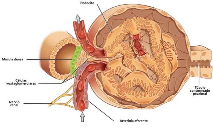
The juxtaglomerular apparatus it is a kidney structure that regulates the functioning of each nephron. Nephrons are the basic structural units of the kidney, responsible for purifying the blood when it passes through these organs.
The juxtaglomerular apparatus is found in the tubule part of the nephron and an afferent arteriole. The tubule of the nephron is also known as the glomerulus, this being the origin of the name to this apparatus.
Article index
In the human kidney there are about two million nephrons that are responsible for the production of urine. It is divided into two parts, the renal corpuscle and the tubule system..
In the renal corpuscle, where the glomerulus is located, the first filtration of the blood takes place. The glomerulus is the functional anatomical unit of the kidney, which is located within the nephrons.
The glomerulus is surrounded by an outer envelope known as Bowman's capsule. This capsule is located in the tubular component of the nephron..
In the glomerulus, the main function of the kidney takes place, which is to filter and purify the blood plasma, as the first stage of urine formation. The glomerulus is actually a network of capillaries dedicated to the filtration of plasma.
The afferent arterioles are those groups of blood vessels responsible for transmitting blood to the nephrons that make up the urinary system. The location of this device is very important for its function, since it allows it to detect the presence of variations in the pressure of the blood that reaches the glomerulus.
The glomerulus in this case, receives blood through an afferent arteriole, and empties into an efferent. The efferent arteriole provides the final filtrate that leaves the nephron, leading to a collecting tube..
Inside these arterioles, a high pressure is produced that ultrafilters the liquids and soluble materials in the blood, being expelled towards the Bowman's capsule. The basic filtering unit of the kidney is made up of the glomerulus and its capsule..
Homeostasis is the ability of living things to maintain a stable internal condition. When there are variations in the pressure received in the glomerulus, the nephrons excrete the hormone renin, to maintain the body's homeostasis.
Renin, also known as angiotensinogenase, is the hormone that controls the body's water and salt balance.
Once the blood is filtered in the renal corpuscle, it passes into the tubular system, where the substances to be absorbed and those to be discarded are selected..
The tubular system has several parts. The proximal contoured tubes are in charge of receiving the filtrate of the glomerulus, where up to 80% of what is filtered in the corpuscles are reabsorbed.
The proximal rectus tubule, also known as the thick descending segment of the loop of Henle, where the reabsorption process is less.
The thin segment of the loop of Henle, which is U-shaped, performs different functions, concentrates fluid content and reduces water permeability. And the last part of the loop of Henle, the distal rectal tube, continues to concentrate the filtrate and ions are reabsorbed.
All this leads to the collecting ducts, which are those that direct urine to the renal pelvis..
Within the juxtaglomerular apparatus we can distinguish three types of cells:
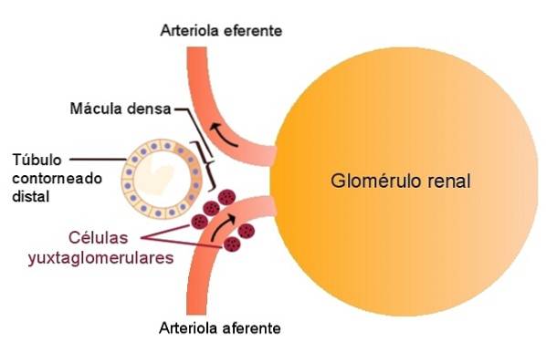
These cells are known by various names, they can be Ruytero cells, granular cells of the juxtagomerular apparatus. They are known as granule cells, because they release granules of renin.
They also synthesize and store renin. Its cytoplasm is riddled with myofibrilia, Golgi apparatus, RER and mitochondria..
For cells to release renin, they have to receive external stimuli. We can categorize them into three different types of stimuli:
The first stimulus that renin secretion provides is that produced by the decrease in blood pressure of the afferent arteriole.
This arteriole is responsible for carrying blood to the glomerulus. This decrease causes a reduction in renal perfusion that, when it happens, causes local baroreceptors to release renin..
If we stimulate the sympathetic system, we also get a response from Ruyter's cells. Beta-1 adrenergic receptors stimulate the sympathetic system, which increases its activity when blood pressure decreases.
As we saw earlier, if the blood pressure drops, renin is released. The afferent arteriole, the one that carries substances, constricts when the activity of the sympathetic system increases. When this constriction occurs, it reduces the effect of blood pressure, which also activates baroreceptors and increases renin secretion..
Finally, another of the stimuli that increase the amount of renin produced are variations in the amount of sodium chloride. These variations are detected by the cells of the macula densa, which increases the secretion of renin..
These stimuli are not produced separately, but all come together to regulate the release of the hormone. But all of them can work independently.
Also known as degranulated cells, these cells are found in the distal convoluted tubule epithelium. They have a tall cubic or low cylindrical shape.
Their nucleus is located inside the cell, they have an infranuclear Golgi apparatus and have spaces in the membrane that allow urine to filter.
These cells, when they notice that the concentration of sodium chloride increases, they produce a compound called adenosine. This compound inhibits renin production, which reduces glomerular filtration rate. This is part of the tubuloglomerular feedback system..
When the amount of sodium chloride increases, the osmolarity of the cells increases. This means that the amount of substances in solution is greater.
To regulate this osmolarity and stay at optimal levels, cells absorb more water, and therefore swell. However, if the levels are very low, the cells activate nitric oxide synthase, which has a vasodilator effect..
Also known as Polkissen or Lacis, they communicate with the intraglomerular ones. They are joined by junctions forming a complex, and are connected to the intraglomerular ones through gap junctions. Gap junctions are those in which the adjoining membranes approach, and the interstitial space between them is reduced.
After many studies, it is still not known with certainty what their function is, but the actions they perform.
They try to connect the macula densa and intraglomerular mesangial cells. In addition, they produce the mesangial matrix. This matrix, made up of collagen and fibronectin, acts as a support for the capillaries.
These cells are also responsible for the production of cytokines and prostaglandins. Cytokines are proteins that regulate cellular activity, while prostaglandins are substances derived from fatty acids..
It is believed that these cells activate the sympathetic system in moments of important discharges, preventing the loss of fluids through the urine, as can happen in the case of a hemorrhage..
After reading so far, we understand that the glomerulus is a network of capillaries in the middle of an artery.
The blood comes through an afferent artery, which divides forming capillaries, which are rejoined to form another efferent artery, which is responsible for the blood outlet. The glomerulus is supported by a matrix formed mainly of collagen. This matrix is called the mesangium.
The entire network of capillaries that make up the glomerulus is surrounded by a layer of flat cells, known as podocytes or visceral epithelial cells. All of this forms the glomerular plume.
The capsule that contains the glomerular tuft is known as Bowman's capsule. It is formed by a flat epithelium that covers it, and a basement membrane. Between Bowman's capsule and the tuft, are parietal epithelial cells and visceral epithelial cells..
The juxtaglomerular apparatus is one formed by:
The interaction of the components of the juxtaglomerular apparatus regulates hermodynamics according to the blood pressure that affects the glomerulus at all times..
It also affects the sympathetic system, hormones, local stimuli, and fluid and electrolyte balance..


Yet No Comments