The caryolysis is one of the changes that are identified in the nuclei of cells when they die as a result of noxa or external damage, such as due to hypoxia (insufficient oxygen supply) or toxic substances.
Its name derives from the Greek karyon, which means "nucleus" and lysis, which can be interpreted as "attenuation" or dissolution "; therefore the term karyolysis literally means "effacement of the nucleus".
This phenomenon occurs during the necrofanerosis stage, such as pyknosis and karyorrhexis, and may be the only nuclear change or be included within a continuum that begins with pyknosis, goes through karyorrhexis and ends in karyolysis..
As in karyorrhexis, the nuclear change precedes the cytoplasmic changes and the process as a whole is accompanied by inflammation of the extracellular matrix, something characteristic of necrosis and that can be considered a fundamental difference with apoptosis, in which no there is the inflammatory complement.
Karyolysis occurs due to the action of nuclear enzymes that in normal conditions help unwind and fragment DNA so that it can be transcribed, but that in conditions of cell death due to noxa (necrosis) begin to disintegrate the nucleus in its entirety.
Article index
The enzymes of the cell nucleus are multiple and very specific, being vital for the physiology of DNA and RNA.
As the genes and chromosomes are structured forming chromatin, it is virtually impossible for DNA transcription and replication to take place, since it is a continuous chain, extremely long and with a very complex three-dimensional spatial conformation..
In order to facilitate the replication and transcription process, nuclear enzymes "cut" the DNA fragment to be transcribed, thus allowing the RNA to be coupled to a linear chain of deoxyribonucleic acid with a very clear start and end..
Also known as "phosphodiesterases", nuclear enzymes are capable of cleaving phosphodiester bonds, key pieces in the structure of nucleic acids, also regulating intracellular levels of cyclic AMP and GMP.
Depending on the site where endonucleases exert their effect, they are classified into two broad categories: nucleases and ligases..
Until now, the effects of nuclease enzymes, responsible for "cutting" the pieces of DNA to allow their replication, have been roughly described, however once the transcription of a DNA fragment has been completed, it must be re-integrated into of the large strand of deoxyribonucleic acid to which it belongs and also to do it in a specific position.
This is where the "ligases" come into play, enzymes capable of "sticking" in its place a DNA chain previously cleaved by phosphodiesterases..
The delicate balance between nucleases and ligases allows the integrity of the genetic material to be maintained, so that when the activity of one enzyme exceeds the other, problems can be predicted.
In order to understand the role of phosphodiesterase in karyolysis, it is essential to know the different types that exist, since they are responsible for the entire process.
In this sense, the ligases have practically no role, in fact their activity is canceled, making it impossible to reverse the process initiated by the nucleases..
Thus, according to the site where they exert their action, nucleases are divided into:
- Endonucleases
- Exonucleases
- Restriction endonucleases
In addition to the enzymes capable of cleaving DNA (also known as DNases), in the nucleus there are also enzymes with the ability to "cut" and model RNA segments, these being known as ribonucleases or RNases..
Although these enzymes are important in the normal physiology of the cell, during the necrosis process they play a secondary role..
Endonucleases are enzymes capable of cutting DNA chains away from their free end, that is, they are capable of separating DNA at any point in the chain.
Endonucleases can cut DNA randomly at any region without matching a particular nucleotide sequence.
Restriction endonucleases are a very special type of endonucleases capable of identifying a specific base sequence in order to cut the DNA chain at that specific point..
They are classified into three groups: Type I, Type II and Type III.
Type I restriction endonucleases require ATP to function (thus consuming energy) and are capable of cleaving up to 1000 base pairs from the recognition sequence.
For its part, the simplest version of restriction endonucleases is Type II; in a process that does not require energy, these enzymes are capable of cutting DNA into variable lengths from the restriction sequence.
Finally, Type III restriction endonucleases, in a process that also consumes energy (ATP), cut the DNA chain into small fragments that do not exceed 25 base pairs from the point of recognition (restriction).
Finally, exonucleases are those enzymes capable of cutting DNA from a free end of the chain, that is, they are specialized enzymes in linear DNA chains previously cleaved by endonucleases..
Thus, the term ENDOnuclease refers to the ability of the enzyme to cut the DNA strand inside (ENDO = inside), while EXOnuclease indicates that the enzyme can only cut the DNA at the free end (EXO = outside).
The synchronized and harmonic activity of all these enzymes allows the complex processes of genetic replication and transcription; However, during necrosis this balance is lost and the DNA begins to fragment until only its free and disorganized basic components remain, which is synonymous with cell death..
Knowing the large number of enzymes present in the nucleus, as well as the way in which they exert their function, it is not difficult to infer the pathophysiology of karyolysis.
Everything begins as a loss of homeostasis between nuclease enzymes and ligases, the effect of the latter being far exceeded by the former; that is, more DNA is destroyed than can be repaired.
In the first instance, endonucleases cut a long DNA chain into small fragments, which are subsequently further reduced by other endonucleases..
Finally, the shorter fragments are lysed from their ends by exonucleases until there are no traces of organized nuclear material, which was enzymatically decomposed..
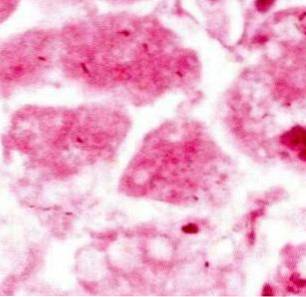
In light microscopy, cells that have undergone karyolysis appear totally pink (eosinophils), making it impossible to identify nuclear material stained purple.
In some cases, an evanescent stain or "ghost" may be seen in the area where the cell nucleus once was, but in general the predominant color will be pink, because there are no more organized nuclear structures capable of capturing hematoxylin..


Yet No Comments