The karyorrhexis It is a phenomenon that occurs in the nucleus of the cell during the process of unscheduled cell death, that is, when the cell dies prematurely due to some type of injury, usually hypoxia (lack of oxygen), toxins or radiation ionizing.
During karyorrhexis, chromatin fragments into small pieces, dispersing within the cell nucleus in a disorganized manner. Due to this, the ability to transcribe DNA is lost..
In conjunction with karyolysis and pyknosis, karyorexis is one of the cytopathological changes present in necrosis.
Previously it was thought that karyorrhexis, karyolysis and pyknosis were three sequential stages of the same process (cell death); however, recent cytopathological studies indicate that these are three separate processes that may or may not overlap.
Karyorrhexis appears during the period of cell death known as necrofanerosis, during which the microscopic changes that precede cell death occur.
To better understand what karyorrhexis is, it is necessary to remember some basic concepts of cell biology.
Article index
Chromatin is the way in which genetic material is organized within the nucleus of the cell when the cell is not replicating.
Most of the time we associate DNA with chromosomes and these in turn to the typical X shape with four more or less elongated arms and a rounded central point..
Although this is true for chromosomes during the active phases of mitosis and meiosis, that is, during cell division, the truth is that in the period known as interface this “typical” configuration does not appear.
Since at the interface the cell does not replicate but rather exerts its physiological functions, it is necessary for the DNA to be more or less accessible to bind with the RNA, and thus start the process of protein synthesis.
If it were in its X configuration, this would be impossible since the DNA strands would be tightly packed with each other, with little or no room for the RNA..
That is why during the interface the DNA "unwinds" forming a more or less chaotic network of fibers known as chromatin..
At the molecular level, chromatin is composed of two fundamental components: Proteins and DNA..
Proteins known as histones are a kind of molecular spool around which the DNA helices are “wound”, in this way a very long DNA strand ends up shortening (by winding) and resembling the beads of a rosary.
Subsequently, each bead (made up of a histone with one and a half turns of DNA) is interwoven with the adjacent ones to further tighten the DNA strands together, so that they are organized into a coherent pattern (chromosome).
The tighter the DNA strands are, it is said that the chromatin is more condensed, on the contrary, when the strands are separated from each other and the DNA chains are looser, it is said that the chromatin is less condensed..
The denser chromatin is known as heterochromatin and these are genes present but not active; On the other hand, lax chromatin is known as euchromatin and corresponds to the DNA segments that are transcribed for the function of a particular cell..
Unlike what happens during apoptosis (programmed cell death) during which a cell that reaches the end of its life becomes a senescent (old) cell and eventually dies without generating inflammation and being replaced by younger cells, during necrosis cell membranes break down initiating a more or less severe inflammatory process.
Although cell death is a process that simultaneously affects both the nucleus and the cytoplasm, the earliest and most obvious changes are at the nuclear level, karyorrhexis being one of them.
In the first instance, due to the release of lytic enzymes, the chromatin begins to fragment. Taking the example in the description of chromatin where the organization of this is compared to the beads of a rosary, when speaking of karyorrhexis it can be said that it is as if the rosary broke into several segments.
This breakdown causes chromatin to disperse and condense into individual, unstructured nuclei, which together take up much more space than organized chromatin in the viable cell..
This increased space required to contain the fragmented chromatin ultimately causes the nuclear membrane to burst, after which the individual chromatin fragments mixed with parts of the nuclear membrane form an amorphous conglomerate in the area where the nucleus of the nucleus would be found. cell.
Once the nucleus "explodes" it is impossible for the cell to fulfill its vital functions, so it dies; This means that when a pathologist observes karyorrhexis in a sample, necrosis (tissue death) is irreversible and all the compromised cells will inexorably die..
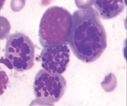
Although karyorrhexis can occur in virtually any cell in the body, it is more common in certain white blood cells (leukocytes), especially basophils and eosinophils..
On the other hand, karyorrhexis is seen with some frequency in cells of the central nervous system, especially in certain tumors such as neuroblastomas..
In the examination of necrotic tissue stained with the hematoxylin-eosin technique and in which karyorrhexis is presented as the main nuclear change associated with cell death, the pathologist and / or cytotechnologist will find characteristic changes that lead to the diagnosis:
The fragmented nuclear material captures a greater amount of hematoxylin, therefore the fragmented and dispersed nucleus appears a more intense purple color.
After karyorrhexis, in the area where the nucleus of the cell would normally be found, dispersed nuclear material is visualized in an amorphous conglomerate that is not surrounded by any type of membrane.
Since the nuclear membrane has been broken, the nuclear material is atomized and dispersed, still having a certain relationship with each other, but in a totally disorganized way and without functional capacity, “floating” free within the cytoplasm.
This finding is unmistakable and synonymous with cell death.


Yet No Comments