It is known as pyknosis to visible changes in the cell nucleus characterized by condensation of chromatin and contraction of the nucleus (becomes smaller) in response to a noxa or cell damage.
In most cases, pyknosis occurs in the stage of necrofanerosis of the cell, being the prelude to its death. Sometimes the only nuclear change during cell death is pyknosis, while in other cases this is just the first step in a series of changes that usually follow the sequence pyknosis -> karyorrhexis -> karyolysis.
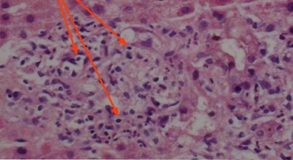
The microscopic examination of the pyknotic nuclei is very characteristic, being these smaller than normal (in relation to normal cells of the same type), and with a greater capacity to capture hematoxylin, for which the pyknotic nucleus usually stains a color more intense blue-purple.
Although pyknosis occurs during necrosis as occurs with karyorrhexis and karyolysis, it can also be seen as part of the normal development of some cells, in response to chronic inflammation and trauma (without necrosis or cell death), as well as in some cases of apoptosis.
In this sense, it is clear that pyknosis can be a pathological process associated with cell death, as well as a normal state of certain cells in response to chromatin condensation..
Article index
For the cell to function properly, the genetic material is dispersed in the nucleus, forming chromatin. The term "dispersed" indicates that the DNA is unwound, forming more or less linear chains in the segments to be transcribed..
The DNA strands that are being transcribed represent the least condensed chromatin, that is, those DNA strands less twisted both on themselves and on the histones.
The DNA segments that should not be transcribed in a specific cell or at any given time "coiled" on themselves in a process known as "condensation" of chromatin. The objective of this process is to save space and keep the genetic material in order..
The less the need for transcription of a given DNA segment, the greater the degree of compaction; thus, during cell division, when there is practically no transcription, chromatin is "squeezed" in its maximum expression to take on the chromosome configuration.
Although it seems a contradiction, in certain cells pyknosis is normal, therefore finding pyknotic nuclei in such cell lines is not synonymous with cell death.
Such is the case with the predecessors of red blood cells known as orthochromatic normoblasts. During this phase of the red blood cell evolution, it is normal for the nucleus to present pyknosis; later in its evolution the cell will expel the nucleus to become a reticulocyte.
Thus, the fact that an orthochromatic normoblast presents pyknosis is something normal and is not related to cell death, on the contrary it is part of its evolution towards maturity.
The same could be said of neutrophils, which during a phase of their maturation present pyknotic nuclei but, far from dying, evolve towards a later stage.
At this stage, the nucleus fragments but does not disperse, so that it could be said that it becomes a "lobed nucleus", this being normal and not associated with cell death..
Something similar occurs with keratinocytes (skin cells), which as they rise along the stratified flat epithelium of which they are part, suffer pyknosis of their nuclei, until finally these disappear in the most superficial layers of the skin. made up mainly of dead cells.
During necrosis there are changes in the permeability of the nuclear membrane, modification of certain molecular signals, and changes in DNA that ultimately induce chromatin condensation..
Unlike what happens under normal conditions, in the cell that dies during necrosis there is no signaling whatsoever that induces protein synthesis and consequently DNA transcription. Therefore, there is no reason for the condensation of chromatin to be reversed, so the genetic material becomes tighter and tighter..
This tight packing is what makes the genetic material take up less space than usual, making the nuclei of cells look smaller (because DNA now takes up less space) and at the same time bluer (there is more concentration acid material that captures hematoxylicin in a smaller space).
Eventually, such tight packaging can cause DNA strands to break apart to give way to karyorrhexis, although this does not always happen; if so, the cell dies with a pyknotic nucleus since it is no longer capable of transcribing DNA.
Unlike karyorrhexis and karyolysis, which occur only in cells that die from necrosis, pyknosis can also be seen in cells that die from apoptosis or "programmed cell death.".
The main difference between necrosis and apoptosis is that during the first process the cell dies prematurely due to an external element (lack of oxygen, toxic, radiation), while in the second the cell reaches its maximum life time and dies.
When pyknosis occurs during apoptosis, the changes are practically the same as those seen in necrosis (condensation of chromatin and contraction of the nucleus), however the changes in the cytoplasm of the cell are different as well as the conditions of the extracellular matrix.
In this sense, during necrosis there is inflammation of the extracellular matrix, while in apoptosis this does not occur..
The technique of sampling and fixing the histopathological or cytopathological material is very important when it is to be examined. Poor technique, a slow process or poor quality of the materials used can induce pyknosis in the tissue once it has been removed from the body.
When this occurs, it is said that a “fixation artifact” has occurred, that is, the nuclei became pyknotic during the processing of the sample and not within the body of the person..
If it is not adequately correlated with the symptoms, the finding of cells with a pyknotic nucleus can lead to false positive diagnoses. If this happens, it is necessary to take and process a new sample in better conditions in order to confirm whether it is a true diagnosis or a false positive..


Yet No Comments