
The secondary lymphoid organs or peripheral are the organs in charge of regulating the cellular interactions of antigens with the cells of the immune system.
That is, in the secondary lymphoid organs the process of recognition of the invading antigen occurs; lymphocytes will be activated only in the presence of non-self.
This ability of lymphocytes to discriminate between what is their own and what is foreign is due to the fact that they have been properly trained in the thymus to do so..
Antigen recognition will lead to a series of events such as phagocytosis, antigen presentation, and activation of other immune cells, with production of antibodies and cytokines..
Due to this function, the secondary lymphoid organs are strategically located in the possible entry doors of antigens to the organism..
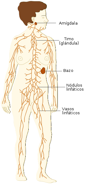
The organs involved are: the lymph nodes and the spleen, which are well-defined capsulated organs, but there are also lymphoid tissues associated with mucous membranes strategically distributed in the body..
The latter are GALT intestinal tissue (Peyer's patches), BALT bronchial tissue, NALT nasopharyngeal tissue (tonsils) and skin (SALT).
Article index
The nodes are complex structures with an ovoid shape, rich in cells of the immune system, especially lymphocytes and macrophages.
Lymph nodes are located in groups throughout the body.
The ganglia are lined by a capsule made up of connective tissue. Trabeculae depart from the capsule that divide the organ into more or less irregular portions.
The capsule is supplied by afferent lymphatic vessels and one more efferent lymphatic vessels converge at the hilum along with the vascular-nervous system of the ganglion..
Inside the ganglion there is an area called the marginal sinus (subcapsular space) from which thin radial channels start, known by their arrangement as radial or intermediate sinuses..
These radial sinuses converge with the efferent lymphatic vessel, at the level of the hilum. As supporting tissue, the ganglion contains reticular cells and connective tissue.
When making a transverse section of the ganglion, two areas of lymphoid tissue are clearly visualized: a cortical area and the medullary area..
Also called thymus-independent area, because this area contains mostly lymphocytes that are not matured in the thymus, that is, B lymphocytes, which are grouped into follicles (primary follicles).
When B cells are activated by the presence of an antigen directly or by contact with an antigen-presenting cell, the B cells become plasma cells..
These activated cells are capable of secreting antibodies and cytokines, in this way the primary follicle becomes a secondary follicle, which is distinguished by the great mitotic activity observed in its central area; Therefore, they are also called Flemming's germ centers..
Memory cells are also formed in this area and other cells such as T lymphocytes and supporting follicular dendritic cells can also be found to a lesser extent..
Also called thymus-dependent area, because here matured lymphocytes are concentrated in the thymus, that is, T lymphocytes..
Despite the clear separation of the two areas, in the independent thymus area, specifically in the deep cortical area, some T lymphocytes may be found, and in the thymus-dependent area (medullary cords) there may also be B lymphocytes or plasma cells..
The function of the ganglia is fundamentally divided into two: the first is the filtration of material from the interstitial fluid and the lymph as these fluids circulate through the canalicular system and reticular cells.
This is how antigens free or bound to antigen presenting cells enter the lymph node through the afferent lymphatic vessels, where they come into contact with the cells of the immune system to be eliminated..
The second function comprises the maintenance of the circulation system of lymphocytes from the blood through the post-capillary venules, where the interaction of lymphocytes with the cells of the vascular elements occurs..
When the ganglia detect an antigen and germinal centers are formed, the ganglion increases significantly in size. This characteristic is easily detectable on palpation in infectious processes..
It is located in the passage of the bloodstream, at the level of the left hypochondrium of the body.
It is an ovoid organ, it is surrounded by a thick fibromuscular capsule, with trabeculae that divide it. In it, two types of tissue are detected: the white pulp and the red pulp..
It is found surrounding the central arteriole, which in turn is protected by a sheath formed mainly by periarteriolar lymphoid tissue.
T lymphocytes surround blood vessels, while B lymphocytes concentrate to form the germinal centers or primary follicles.
Macrophages are found on the border between the white and red pulp zones, which act as antigen-presenting cells and engulf damaged cells..
The red pulp surrounds the white pulp and is mostly made up of erythrocytes and around the vessels are B lymphocytes.
It is supplied by vascular sinusoids that connect with the splenic vein.
The spleen filters half of the body's blood volume every day, being an effective mechanism to clean the blood of any invading microorganism that may have entered the circulation, in addition to eliminating aging or non-functional cells.
Therefore, the spleen fulfills two types of functions, one related to the immune system and the other non-immunological..
The non-immunological ones include the maintenance of homeostasis, removing damaged erythrocytes from the circulatory system, converting hemoglobin into bilirubin, and releasing iron for reuse..
While the immune function is related to facilitating the immune response, both humoral and cellular, since it contains mature lymphocytes and plasma cells.
These specialized tissues are distributed in the body and present characteristic cells of the place with different functions, but all have lymphocytes in their composition..
Generally specialized tissues take up cell-bound antigens.
Mucosa-associated lymphoid tissue is organized into primary and secondary follicles as described in the lymph nodes and spleen, rich in B lymphocytes and plasma cells respectively..
Around the follicles are intraepithelial lymphocytes, which mostly correspond to the CD8 or cytotoxic type, which interact directly with the antigen.
At these sites, the immune response is reinforced by the action of IgA-type antibodies, normally present in the mucous membranes..


Yet No Comments