The lung segments They are portions or divisions of the lungs with specific characteristics and functions. This division occurs thanks to the bronchial bifurcation.
The pulmonary segments can also be considered macroscopic units of the lung, provided with a bronchus (segmental bronchus) and a complementary branch of the pulmonary artery.
Each of these segments has between 30 and 60 bronchioles, a particular venous and arterial supply, so they function independently.
They cannot be seen from the outside of the lung, but doctors must use X-rays or bronchoscopy, and are separated by connective tissue septa.
The images achieved to detect these segments in the lung are usually incomplete or confusing..
The lung segments serve to guide thoracic surgeons in interpreting images of the lungs and in surgical procedures.
The right lung is divided into 3 lobes (upper, middle and lower), divided with 2 fissures, while the left has 2 lobes with a fissure.
For the interpretation of two-dimensional images, scientists created a classification system for vessels and bronchi in 5 lobe regions, by detecting edge and curved surface, taking advantage of the linear appearance of fissures in the lobes..
In addition, there is a kind of global lung atlas that serves as a template and is coded. Regarding three-dimensional images, the methods use the Gaussian approach and the analysis of Hessian matrices..
It is worth saying that a weakness of these systems based on anatomical knowledge is that they ignore individual variability, which could lead to segmentation failures when the methods are applied to the "new" exams..
Whereas if the focus is based on shapes in the image space, these risks of errors decrease.
Lung segmentation is normally done based on the location and direction of the main and segmental bronchi.
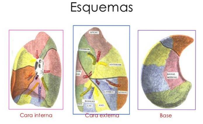
In the right lung there are 10 segments:
In the left lung there are also 10 segments, but since there are cases in which they are ventilated through the same bronchus, two are counted as one.
It should also be clarified that there are a couple of marked differences with respect to the segments of the right lung: the combination of segments and the presence of a “lingular lobe” incorporated in the upper lobe of the left lung, which replaces the middle lobe..
It should be noted that there is a nomenclature to name the elements of each lung segment (segmental bronchus, artery and veins).
The rules of this nomenclature dictate that a capital letter must be used (S, B, A or V, depending on whether it refers to segments, lobar bronchi, arteries or vein), followed by a number, which indicates to which segment the element belongs in question.
That is, the name of the element already illuminates its location in the lung. For example: B2 refers to the bronchus that ventilates segment 2.
They are the bronchi that carry air to the lung segments.
The B1, B2 and B3 are those that ventilate the segments that make up the upper lobe of the lung, while B4 and B5 ventilate the middle lobe.
The lower lobe of the right lung is ventilated by bronchi B6 to B10
This lobe is the area with the most bronchi, because it is also the area with the most volume and lung parenchyma, which is why it is the part that needs more ventilation..
In the case of the bronchi that ventilate the left lung, from B1 to B5 they go to the upper lobe; B7 and B8 ventilate the anteromedial basal segment and from B6 to B10 they go to the lower lobe segments.


Yet No Comments