
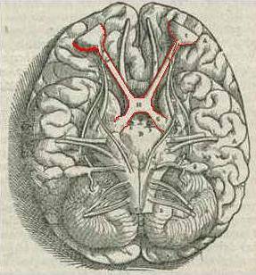
The optic chiasma It is a brain structure in which the fibers of the optic nerves partially intersect. That is, it is a region of the brain that acts as a junction point between the optic nerve of the right eye and the optic nerve of the left eye..
This narrowing is located in the anterior cerebral fossa, located just in front of the sella turcica. It is about twelve millimeters wide, eight millimeters long and about four millimeters high..
The main function of this area of the brain is to integrate and unify the visual stimuli captured through the eyes, with the aim of generating informational elements that can be sent to other regions of the brain..
Likewise, the optic chiasm performs the particular function of crossing the fibers of the optic nerves, so the right region of the chiasm processes the left eye and the left region processes the right eye.
Article index
Optic chiasm is a term that comes from the Greek and means cross arrangement. Biologically, this word refers to a small brain region.
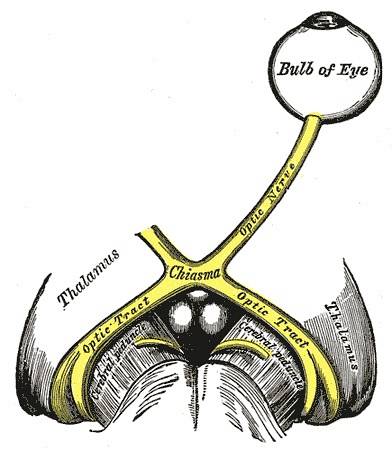
The optic chiasm is a structure of the brain that is characterized by being a point of attachment of the axonal fibers of the optic nerves. That is, it is the area of the brain where the visual stimuli captured by the right eye and the left eye end up..
In the optic chiasm, the axonal fibers of the optic nerves intersect. At this junction, half of the fibers pass from the right optic nerve to the left optic tract and from the left optic nerve to the right optic tract.

In this sense, the optic chiasm is a structure that allows visual information to intersect and connect the optic nerves with the optic tracts..
The main peculiarity of the optic chiasm is that it is not only a point of union between the two optic nerves, but also, it is the point at which the optic fibers of these nerves partially cross.
In this way, the optic chiasm is an essential brain structure for processing visual information. This region is observed in all vertebrates, including cyclostomes.
The optic chiasm is itself a nerve structure. It has a shape similar to the Greek letter chi and is characterized by deriving from the fusion of the two optic nerves.
The structure of the optic chiasm arises through the axonal fibers of each optic nerve and continues posteriorly with the two optic strips.
The optic chiasm is a small brain structure. It is approximately 12-18 millimeters wide, about eight millimeters long, and about four millimeters high..
Just above the optic chiasm is the floor of the third ventricle, a structure with which it is directly interrelated. Laterally, the optic chiasm establishes connection with the internal carotid arteries and, inferiorly, with the sella turcica and the pituitary gland..
The optic chiasm is a brain region that plays an important role in the optic pathway. That is, it constitutes a structure that is essential to transmit and integrate visual information and, therefore, allow vision as a perceptual sense..
The optic pathway is therefore a set of brain structures that is responsible for transmitting nerve impulses from the retina to the cerebral cortex. This process is done through the optic nerve.
The receptor cells of the optic nerve are the rods and cones, which transform the images received into nerve impulses that are transferred to the brain and conducted by different structures..
In this sense, the role of the optic chiasm can divide the optic pathway into two main categories: structures anterior to the optic chiasm and structures posterior to the optic chiasm..
Before the perceived information reaches the brain region of the optic chiasm, a main structure for the perception of visual stimuli participates in the optic pathway: the optic nerve..
The optic nerve is formed by the axons of the ganglion cells of the retina of the eye. These nerves are covered by meninges, begin in the posterior scleral foramen and end in the optic chiasm itself.
The optic nerve has a variable length of between four and five centimeters approximately, and is characterized by being divided into four main portions:
Once the information is transmitted from the optic nerves to the optic chiasm, and the latter has integrated and interlaced the visual stimuli, the information is directed to other brain regions.
Specifically, posterior to the optic chiasm, the optic pathway has four areas: the optic strips, the external geniculate body, Gratiolet's optic radiations and the visual areas..
Optic streaks originate in the region immediately posterior to the chiasm. Each band is separated from the other through the pituitary stalk in the lower part and through the third ventricle in the upper region.
The optic tracts contain the nerve fibers that come from the temporal retina and the nasal retinas. In this region there is a new arrangement of nerve fibers. Most of the fibers of the girdle end at the level of the geniculate body and a small percentage is directed towards the superior cudrigémic tubercle.
The external geniculate body is the next structure of the optic pathway. This region generates a connection of the axons of the ganglion cells with the neurons inside them..
The synapse between cells and neurons is responsible for coding in a certain part the nerve signals, elaborating the visual information.
Finally, the neurons of the external geniculate body extend their axons through optical radiation, which continue to form the external wall of the lateral ventricles..
Certain fibers surround the ventricles establishing relationships with the internal capsule and forming the loop of Myere. Instead, most of the fibers are directed toward Brodman's area 17 of the cerebral cortex..
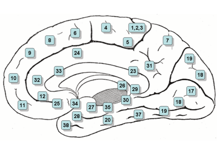
Finally, the transmission of the visual nerves ends in the visual areas, which are formed by areas 17, 18 and 19 of Brodman.
Of all of them, area 17 is the main visual region, which is located at the level of the interhemispheric cleft, on the posterior surface of the occipital cortex of the brain..
Brodman's area 17 is divided into two parts by the calcarine fissure, so the region of the cortex next to this region is called the calcarine cortex.
Brodman's areas 18 and 19, on the other hand, are brain association regions. They establish interhemispheric connections in which the visual information that arrives through the optical pathway is analyzed, identified and interpreted.
Lesions in the optic chiasm are quite infrequent, thus being one of the regions of the optic pathways that are less frequently damaged..
The optic chiasm is located inside the skull and in the lower region of the brain, so it rarely suffers from severe injury. In fact, few cases of lesions in the optic chiasm have been detected today. However, certain types of hemianopia can arise due to damage to this brain region..
Hemianopia is a pathology that involves lack of vision or blindness and is characterized by affecting only half of the visual field. At present, different types of hemianopia have been detected, of which only two respond to damage to the optic chiasm: binasal hemianopia and bitemporal hemianopia..
Binasal hemianopia is a type of heteronymous hemianopia that affects the left half of the visual field of the right eye and the right half of the left visual field, and is caused by a lesion in the optic chiasm.
For its part, bitemporal hemianopia is characterized by affecting the right half of the visual field of the right eye and the left half of the visual field of the left eye, and is also due to a lesion in the optic chiasm that is sometimes caused by a pituitary tumor.


Yet No Comments