The chytridomycetes they are fungi that generally present simple monoflagellate zoospores, with the flagella inserted posteriorly. They present alternation of generations with a sexual and an asexual phase.
They are ubiquitous organisms, they are found, both in the tropics and in cold regions, in the soil, fresh water or in saline estuaries. Most species are parasitic on vascular plants, rotifers, phytoplankton, bryophytes, and other fungi, including other chytridomycetes..
Some of these fungi are saprophytes. There are some anaerobic species that inhabit well-defined regions of the digestive tract of herbivorous mammals..
A chytridomycete, Batrachochytrium dendrobatidis, it is the agent responsible for a life-threatening disease that affects amphibian animals. This disease is called chytridiomycosis. It has caused mass mortalities, population declines, and extinctions of amphibian populations and species around the world..
Article index
Chytridomycetes show alternation of generation. The somatic phase has a variable shape. It can present as an isolated cell, an elongated hypha, or a well-developed non-septate (coenocytic) mycelium, depending on the species. They have spores with flagella. The flagella are simple, without comb-like fibrils (mastigonemes).
Zoospores are produced in a thin-walled sporangium. These zoospores are mobile, driven by a single flagellum inserted posteriorly. The flagellum is shaped like a whip. In some species the zoospore shows a set of honeycomb-like tubular membranes (rumposome).
Cell walls contain chitin and glucan. The thallus can produce one or more sporangia on a network of rhizoids. If it is a single sporangium, the thallus is called monocentric. If there are several, it is called polycentric. They are usually microscopic.
Chytridiomycetes is a class of fungi located within the phylum Chytridiomycota. This phyllum was also contained by the classes Blastocladiomycota and Neocallimastigomycota.
Studies based on zoospore ultrastructure and morphological characteristics suggested that the group was monophyletic. Molecular studies and multilocus data, however, showed that the phyllum was actually polyphyletic or paraphyletic, suggesting that Blastocladiomycota and Neocallimastigomycota actually formed sister clades..
Because of this, these two taxa were elevated to the phylum level. The remaining Chytridiomycota have then been divided into five classes. The Chytridiomycetes class is the most diverse in terms of number of species.
Chytridomycetes show alternation of generations. One generation has haploid gametothals and another has diploid sporothals. Gametotali develop male and female gametangia. Gamentangia will produce mobile gametes called planogametes.
A male and female gamete fuse in the middle to form a biflagellate zygote that later loses the flagella and becomes encyst. The germination of the diploid cyst will produce a sporothal. Upon maturation, the sporothal will develop zoosporangia of two types: mitosporangia and meiosporangia..
Mitosporangia have a thin, colorless wall. Inside they will produce diploid zoospores by mitotic division. The zoospores are released, swim for a time, encyst and germinate to originate new diploid sporothals.
Meiosporangia have thick, pigmented cell walls. These will produce, by meiosis, haploid zoospores. These spores, known as dormancy zoospores, encyst and will later germinate to form new gametotali..
Chytridomycetes can be saprophytes, breaking down refractory materials, such as pollen, cellulose, chitin, and keratin. These fungi release chemicals that degrade these materials and subsequently acquire the nutrients through the rhizoids..
Anaerobic species feed by digestion of the plant cell wall of the rumen of herbivorous mammals. These organisms produce large amounts of extracellular cellulases.
These enzymes can interact with those produced by other microorganisms. Studies indicate that chytridomycetes play an important role in ruminal digestion.
Parasitic chytridomycetes feed on tissues or nutrients from their hosts, which may be plants, animals, or other fungi, including other chytridomycetes..
Asexual reproduction occurs in diploid organisms, or sporothals. These will produce two types of zoospores: mitotic and meiotic..
Mitotic zoospores are produced in mitotic reproductive sporangia (mitosporangia). These when germinating produce new sporothals.
Meiotic zoospores are produced in meiosporangia. These zoospores, when germinating, produce haploid gametotali.
Sexual reproduction occurs in the haploid thalli or gametotali. These thalli will produce, by mitosis, male and female mobile sexual gametes (planogametes). Planogametes fuse producing a diploid spore that, when germinating, will give rise to a sporothal..
Among the plant pathogenic Chitridomycetes, mention may be made of Olpidium brassicae. This species is an obligate parasite of plants such as clovers and cabbages. Its greatest danger is represented by the fact that it acts as a vector for many necroviruses.
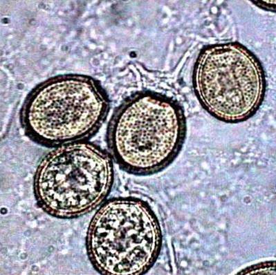
The disease known as potato black wart, is caused by a chytidromycete called Synchytrium endobioticum. The fungus produces dormant spores. Dormant spores, when germinating, produce zoospores.
These infect plant cells, producing a thallus, or sometimes a zoosporangium, which causes infection. The government of the United States of America considers this species as a phytopathogen of possible use in bioterrorism.
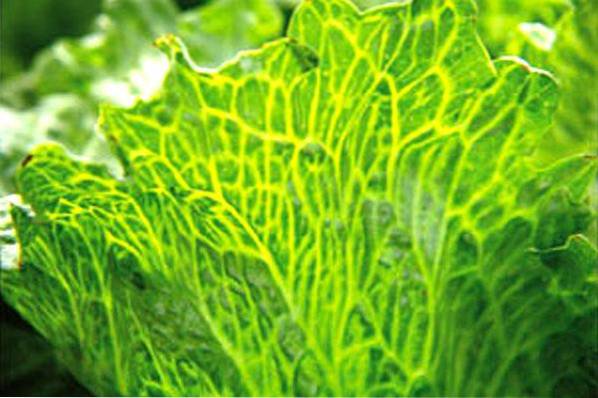
Physoderma maydis it is a chytridomycete responsible for the disease known as maize brown spot. The first symptoms of the disease appear on the leaves.
These consist of small chlorotic spots arranged in the form of alternating bands of healthy and diseased tissue. As the disease progresses, the bands also appear on the stem. Eventually the bands come together and cause stem rot.
Chytridiomycosis, produced by Batrachochytrium dendrobatidis, it is perhaps the most important disease caused by chytridomycetes in animals. This fungus, discovered and described at the end of the 20th century, is considered an emerging pathogen.
It has been documented in numerous amphibian species and in increasingly wide geographic regions. It has caused drastic declines in amphibian populations, and even local extinctions.
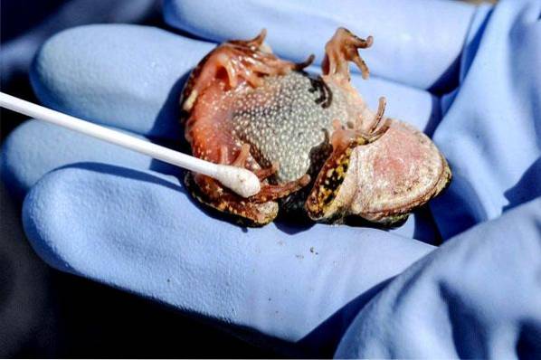
Batrachochytrium dendrobatidis it lodges in the skin cells of infected amphibians. The pathological abnormality due to chytridomycete consists of a thickening of the outer layer of the skin. No other alteration has been found in the internal organs.
It has been hypothesized that B. dendrobatidis disrupts the normal regulatory functioning of the skin of diseased amphibians. Electrolyte depletion and osmotic imbalance that occurs in amphibians due to severe episodes of chytridiomycosis would be enough to cause death..


Yet No Comments