The consensual reflex it is any reflex observed on one side of the body when the other side has been stimulated. This reflex is mainly evidenced in the process of contraction of the pupil of both eyes when illuminating only one of them (Dictionary, 2017).
The pupillary response to light is the reduction in the size of the pupil when illuminating the eye directly. This is the most common stimulus applied for the contraction of the hole located in the center of the iris..
The process of contraction of both pupils in a uniform way when the stimulus is generated in a single eye is known as a consensual reflex (Backhaus, 2011).
The consensual reflex is important in determining whether there is neurological or central nervous system damage. If the contraction of the pupils occurs unevenly, it can be concluded that there is damage to the cranial nerves of the patient. Similarly, the consensual reflex can help determine if there is damage to the retina or oculomotor nerves..
There are several tests and light stimuli that can be used to demonstrate the normal reaction of the consensual reflex in both pupils. These tests include the gradual lighting of a room, the direct application of light to one of the two eyes, or the oscillating light test..
The consensual reflex is different from the photomotor reflex, the latter being the one that takes place in the eye in which the light stimulus is applied directly and whose effect is also the contraction of the pupil.
Article index
The size of the pupil is determined by the interaction of the sympathetic and parasympathetic nervous systems, which are connected to the iris.
These systems are controlled by the central nervous system, which sends signals to the brain influenced by numerous factors, such as lighting, observation distance, state of vigilance and cognitive state (Dragoi, 1997).
The reduction in pupil size occurs when the circular muscle of the eye, controlled by the sympathetic nervous system, contracts in response to an external stimulus of light..
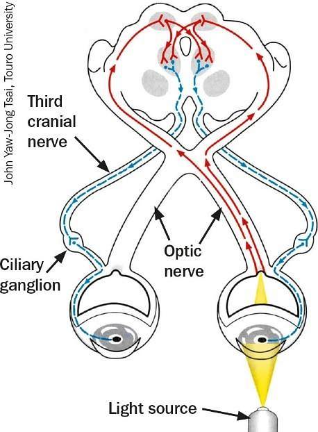
Pupillary constriction of each eye occurs when the retina, or optic nerve, and the pretectal nucleus of each eye take sensory information from outside.
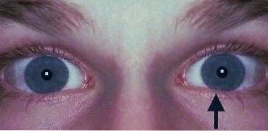
When one of an individual's eyes is covered and the other eye is illuminated, the contraction of the pupil of both eyes must occur simultaneously and uniformly..
This happens thanks to the sending of an afferent signal through the optic nerve that connects with the Edinger-Westphal nucleus, whose axons cross the oculomotor nerves of both eyes..
The size and shape of the pupil in normal light conditions is in a range of 1 to 8 millimeters. When the pupils work correctly, they are said to be isochoric, this means that they react in the same way to the light stimulus. When this stimulus is modified, the pupils must grow symmetrically and simultaneously.
To assess that the pupils are functioning normally, a consensual reflex test is usually applied..
This test consists of illuminating both eyes independently, in such a way that a direct pupillary response is produced in the eye that is being illuminated and an indirect response in the eye that is not receiving is stimulus..
If the optic nerve of the illuminated eye is damaged, the pupil reflex does not take place, therefore, the consensual reflex does not take place, since the eye that is not being stimulated does not receive any message.
However, if the optic nerve of the eye that is being illuminated and the oculomotor nerve of the eye that is not being stimulated are in perfect condition, the consensual reflex will take place, since the signal can be sent by one eye and received by the other. (Bell, Wagoner, & Boyd, 1993).
There are some disorders that can occur in the nervous system of the eye that can affect the process of contraction of the pupil.
These disorders can affect the parasympathetic system and cause the consensual response to light to take place irregularly (Levatin, 1959). Some of these disorders may include the following:
1-Inflammation of the optic nerve (optic neuritis).
2-High intraocular pressure (severe glaucoma).
3-Direct or indirect ocular trauma (traumatic optic neuropathy).
4-Tumor of the optic nerve.
5-Disease in the eye socket.
6-Optic atrophy.
7-Infections or inflammations of the optic nerve.
8-Retinal diseases
9-Intracranial pathologies
10-Brain injuries
11-Pharmacological blocks (Lowth, 2017)
The oscillating light test is used to detect the presence of reactive pupillary afferent defects. This means that the test is used to determine if there is any difference in the way both eyes respond to the application of light on one of the two eyes..
The test is quite useful to detect diseases of the retina or optic nerve that cause the pupils to contract asymmetrically (Broadway, 2012).
The steps to carry out this test are as follows:
1-Use a flashlight that can be focused close to the eye in a dimly lit room.
2-Ask the patient to look into the distance while the eye lights up. This will prevent the pupil from contracting due to the reaction to the proximity of the flashlight during the test..
3-Move the flashlight deliberately from one eye to another, illuminating each eye independently. Be careful not to move the flashlight close to the nose, as this may stimulate the pupil's response to a nearby object..
4-Keep moving the flashlight the same distance from each eye to ensure that each eye is receiving the same stimulus.
5-Hold the flashlight for three seconds in each eye, allowing the movement of the pupil to stabilize. Observe what happens to the other pupil during this process.
6-Repeat the test several times in order to identify what happens to the pupil of each eye when it is illuminated.


Yet No Comments