
The endoplasmic reticulum it is a membranous cellular organelle present in all eukaryotic cells. This complex system occupies approximately more than half of the membranes in a common animal cell. The membranes continue until they meet the nuclear membrane, forming a continuous element.
This structure is distributed throughout the cell cytoplasm in the form of a labyrinth. It is a kind of network of tubules connected to each other with sac-like structures. Within the endoplasmic reticulum, protein and lipid biosynthesis occurs. Almost all proteins that must be taken to the outside of the cell pass through the reticulum first.
The reticulum membrane is not only responsible for separating the interior of this organelle from the cytoplasmic space and mediating the transport of molecules between these cell compartments; It is also involved in the synthesis of lipids, which will form part of the plasma membrane of the cell and the membranes of the other organelles..
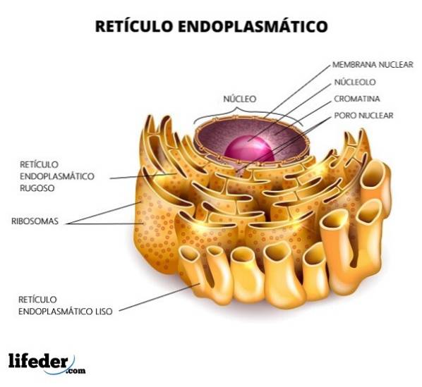
The reticulum is divided into smooth and rough, depending on the presence or absence of ribosomes in its membranes. The rough endoplasmic reticulum has ribosomes attached to the membrane (the presence of ribosomes gives it a “rough” appearance) and the shape of the tubules is slightly straight.
For its part, the smooth endoplasmic reticulum lacks ribosomes and the shape of the structure is much more irregular. The function of the rough endoplasmic reticulum is directed mainly to the processing of proteins. In contrast, smooth is responsible for lipid metabolism.
Article index
The endoplasmic reticulum is a membranous network present in all eukaryotic cells. It is composed of saccules or cisterns and tubular structures that form a continuum with the membrane of the nucleus and are distributed throughout the cell.
The reticulum lumen is characterized by high concentrations of calcium ions, in addition to an oxidizing environment. Both properties allow it to fulfill its functions.
The endoplasmic reticulum is considered the largest organelle present in cells. The cell volume of this compartment covers approximately 10% of the cell interior.
The rough endoplasmic reticulum has a high density of ribosomes on the surface. It is the region where all the processes related to protein synthesis and modification occur. Its appearance is mainly tubular.
The smooth endoplasmic reticulum does not have ribosomes. It is abundant in cell types that have an active metabolism in lipid synthesis; for example, in the cells of the testes and ovaries, which are steroid-producing cells.
Similarly, the smooth endoplasmic reticulum is found in a fairly high proportion in liver cells (hepatocytes). The production of lipoproteins occurs in this area..
Compared with the rough endoplasmic reticulum, its structure is more complicated. The abundance of the smooth versus the rough reticulum depends primarily on the cell type and its function..
The physical architecture of the endoplasmic reticulum is a continuous membrane system made up of interconnected sacs and tubules. These membranes extend to the core, forming a single lumen.
The lattice is built by multiple domains. The distribution is associated with other organelles, with different proteins and with the components of the cytoskeleton. These interactions are dynamic.
Structurally, the endoplasmic reticulum consists of the nuclear envelope and the peripheral endoplasmic reticulum, made up of the tubules and sacs. Each structure is related to a specific function.
The nuclear envelope, like all biological membranes, is made up of a lipid bilayer. The interior delimited by this is shared with the peripheral reticulum.
The sacs that make up the endoplasmic reticulum are flat and often stacked. They contain curved regions at the edges of the membranes. The tubular network is not a static entity; can grow and restructure.
The sac and tubule system is present in all eukaryotic cells. However, it varies in shape and structure depending on the cell type..
The reticulum of cells with important functions in protein synthesis is composed primarily of sacs, while the cells most related to lipid synthesis and calcium signaling are composed of a greater number of tubules..
Examples of cells with a high number of sacs are the secretory cells of the pancreas and B cells. In contrast, muscle cells and liver cells have a network of prominent tubules..
The endoplasmic reticulum is involved in a number of processes including protein synthesis, trafficking and folding, and modifications, such as disulfide bond formation, glycosylation, and the addition of glycolipids. In addition, it participates in the biosynthesis of membrane lipids.
Recent studies have linked the reticulum with responses to cellular stress, and may even induce apoptosis processes, although the mechanisms have not been fully elucidated. All these processes are described in detail below:
The endoplasmic reticulum is closely linked to protein trafficking; specifically to proteins that must be sent abroad, to the Golgi apparatus, to lysosomes, to the plasma membrane and, logically, to those that belong to the same endoplasmic reticulum.
The endoplasmic reticulum is the cellular behavior involved in the synthesis of proteins that must be carried outside the cell. This function was elucidated by a group of researchers in the 1960s, studying cells of the pancreas whose function is to secrete digestive enzymes..
This group, led by George Palade, managed to label proteins using radioactive amino acids. In this way it was possible to trace and locate the proteins by a technique called autoradiography..
Radioactively labeled proteins could be traced to the endoplasmic reticulum. This result indicates that the reticulum is involved in the synthesis of proteins whose final destination is secretion..
Later, the proteins move to the Golgi apparatus, where they are "packed" into vesicles whose content will be secreted..
The secretion process occurs because the membrane of the vesicles can fuse with the plasma membrane of the cell (both are lipid in nature). In this way, the content can be released to the outside of the cell..
In other words, secreted proteins (and also lysosome and plasma membrane targeting proteins) must follow a specific pathway that involves the rough endoplasmic reticulum, the Golgi apparatus, secretory vesicles, and finally the exterior of the cell..
Proteins that are destined to be incorporated into some biomembrane (plasma membrane, Golgi membrane, lysosome, or reticulum) are first inserted into the reticulum membrane and are not released instantly into the lumen. They must follow the same route for secretory proteins.
These proteins can be located within the membranes by a hydrophobic sector. This region has a series of 20 to 25 hydrobophic amino acids, which can interact with the carbon chains of phospholipids. However, the way in which these proteins insert is variable..
Many proteins cross the membrane only once, while others do so repeatedly. Likewise, it may in some cases be the end of the carboxyl terminal or the amino terminal.
The orientation of said protein is established while the peptide grows and is transferred to the endoplasmic reticulum. All protein domains that point towards the reticulum lumen will be found on the outside of the cell in their final location..
Molecules of a protein nature have a three-dimensional conformation necessary to carry out all their functions..
DNA (deoxyribonucleic acid), by a process called transcription, passes its information to an RNA (ribonucleic acid) molecule. The RNA then passes into the proteins through the process of translation. Peptides are transferred to the reticulum when the translation process is in progress.
These chains of amino acids are arranged in a three-dimensional way within the reticulum with the help of proteins called chaperones: a protein of the Hsp70 family (heat shock proteins or heat shock proteins for its acronym in English; the number 70 refers to its atomic mass, 70 KDa) called BiP.
The BiP protein can bind to the polypeptide chain and mediate its folding. Likewise, it participates in the assembly of the different subunits that make up the quaternary structure of proteins..
Proteins that have not been correctly folded are retained by the reticulum and remain bound to BiP, or become degraded.
When the cell is subjected to stress conditions, the reticulum reacts to it and, as a consequence, the correct folding of proteins does not occur. The cell can turn to other systems and produce proteins that maintain reticulum homeostasis.
A disulfide bridge is a covalent bond between the sulfhydryl groups that are part of the amino acid structure cysteine. This interaction is crucial for the functioning of certain proteins; likewise, it defines the structure of the proteins that present them.
These bonds cannot be formed in other cell compartments (for example, in the cytosol), because it does not have an oxidizing environment that favors its formation..
There is an enzyme involved in the formation (and breaking) of these bonds: the protein disulfide isomerase.
In the reticulum, the glycosylation process occurs, at specific asparagine residues. Like protein folding, glycosylation occurs while the translation process is running.
The oligosaccharide units are made up of fourteen sugar residues. They are transferred to asparagine by an enzyme called oligosacaryltransferase, located in the membrane.
While the protein is in the reticulum, three glucose residues and one mannose residue are removed. These proteins are taken to the Golgi apparatus for further processing..
On the other hand, certain proteins are not anchored to the plasma membrane by a portion of hydrophobic peptides. In contrast, they are attached to certain glycolipids that function as an anchoring system and are called glycosylphosphatidylinositol (abbreviated as GPI)..
This system is assembled in the reticulum membrane and involves the binding of GPI at the terminal carbon of the protein..
The endoplasmic reticulum plays a crucial role in lipid biosynthesis; specifically, the smooth endoplasmic reticulum. Lipids are an indispensable component of the plasma membranes of cells.
Lipids are highly hydrophobic molecules, so they cannot be synthesized in aqueous environments. Therefore, its synthesis occurs in association with already existing membranous components. The transport of these lipids occurs in vesicles or by transporter proteins.
The membranes of eukaryotic cells are made up of three types of lipids: phospholipids, glycolipids, and cholesterol..
Phospholipids are derived from glycerol and are the most important structural constituents. These are synthesized in the region of the reticulum membrane that points to the cytosolic face. Different enzymes participate in the process.
The membrane grows by the integration of new lipids. Thanks to the existence of the enzyme flipase, growth can occur in both halves of the membrane. This enzyme is responsible for transferring lipids from one side of the bilayer to the other..
The processes of synthesis of cholesterol and ceramides also occur in the reticulum. The latter travels to the Golgi apparatus to produce glycolipids or sphingomyelin.
The calcium molecule participates as a signaling agent in different processes, be it the fusion or association of proteins with other proteins or with nucleic acids..
The interior of the endoplasmic reticulum has calcium concentrations of 100-800 uM. Calcium channels and receptors that release calcium are found in the reticulum. Calcium release occurs when phospholipase C is stimulated by the activation of G-protein-coupled receptors (GPCRs).
In addition, the elimination of phosphatidylinositol 4,5 bisphosphate in diacylglycerol and inositol triphosphate occurs; the latter is responsible for the release of calcium.
Muscle cells possess an endoplasmic reticulum specialized in the sequestration of calcium ions, called the sarcoplasmic reticulum. It is involved in the processes of muscle contraction and relaxation.


Yet No Comments