The sack of douglas or rectouterine bag is an extension of the peritoneum and is connected anteriorly with the uterus and the posterior fornix of the vagina and posteriorly with the rectum in women. The term was derived from the Scottish anatomist James Douglas, who conducted studies focused on the female anatomy..
This sac is of great clinical importance. The floor of this structure is only 5.5 cm from the anus. When performing a rectal or vaginal exam, any abnormality can be easily felt.
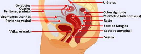
Being the most dependent part, pus, blood, or ascitic fluids tend to accumulate there; these fluids can be drained or samples can be collected from the posterior fornix of the vagina.
The peritoneum descends from the anterior abdominal wall toward the upper surface of the urinary bladder. Then it travels directly over the anterior surface of the uterus.
From there it travels downward and covers the upper part of the posterior surface of the vagina, where it forms the anterior wall together with the sac of Douglas..
Article index
Eight ligaments hold the uterus in its normal position by anchoring it to the pelvic cavity. Some of these ligaments are actually extensions of the parietal peritoneum in different directions:
Pelvic inflammatory disease is an infection in the female reproductive organs. This is one of the most serious complications of sexually transmitted diseases in women..
It can lead to irreversible damage to the uterus, ovaries, fallopian tubes, among other parts of the female reproductive system. It is also known as the leading cause of infertility in women.
Pelvic inflammatory disease occurs when disease-causing organisms travel from the cervix to the upper genital tract. Symptoms are usually pain in the lower abdomen along with back pain.
In order to diagnose the cause, doctors generally order cervical sweeps and also a collection of fluids from the sac of Douglas. The disease is usually treated with antibiotics and fluids begin to be absorbed over time..
A pocket of pus during an infection of the fallopian tubes and ovaries is known as an ovarian abscess. These can develop in women who have pelvic inflammatory disease. The fluids generated by this infection tend to be collected in the Douglas sac.
These abscesses are diagnosed with physical exams or ultrasound. Treatment is usually with antibiotics, but if the infection persists, the abscess has to be drained..
Drainage is done with a long needle that cuts the abscess during a laparoscopy or laparotomy. Sometimes the entire infected tube has to be surgically removed.
Hydatidiform moles are a rare mass or growth inside the uterus in early gestation. This is the result of a lot of tissue production that should become the placenta. In these cases there are inflammatory processes with fluids that can be observed in the sac of Douglas.
A pelvic exam might show signs similar to a normal pregnancy. The size of the uterus may be abnormal and there may be no heartbeat from the pregnant baby. Vaginal bleeding may occur. In these cases, a dilation and curettage is recommended to treat hydatidiform moles.
This type of pregnancy occurs when the fertilized egg implants in the fallopian tubes or elsewhere in the abdomen. In these cases the pregnancy cannot continue and emergency treatment is required. Symptoms include but are not limited to mild vaginal bleeding and pelvic pain.
The women most at risk of presenting this type of pregnancy are those who have had pelvic inflammatory diseases in the past, with implications for the sac of Douglas.
If left untreated, this abnormality can lead to a ruptured fallopian tube with severe internal bleeding..
Endometriosis is an abnormality in which the endometrium, which is the tissue that normally lines the inside of the uterus, begins to grow outside the uterus. This tissue that is not in place, also bleeds when the woman menstruates and some of that blood can be collected in the sac of Douglas..
This can lead to a secondary complication known as dysneuria, in which it becomes painful for the woman to have sexual intercourse..
Peritonitis is an inflammation of the peritoneum and is usually caused by a bacterial or fungal infection. If left untreated, the infection can quickly spread into the blood and other organs causing systemic failure of all organs and death. This disease causes excessive fluid that can be collected in the sac of Douglas..
Cysts on the ovaries are fluid-filled sacs within or on the surface of the ovaries. Many cysts go unnoticed, but others can rupture causing serious complications. The fluid product of the rupture accumulates in the sac of Douglas.


Yet No Comments