
A sarcomere or sarcomere is the fundamental functional unit of skeletal muscle, that is, of skeletal and cardiac muscle. Skeletal muscle is the type of muscle that is used in voluntary movement and cardiac muscle is the muscle that is part of the heart..
Saying that the sarcomere is the functional unit means that all the components necessary for contraction are contained in each sarcomere. In fact, skeletal muscle is made up of millions of tiny sarcomeres that shorten, individually, with each muscle contraction..
Herein lies the main purpose of the sarcomere. Sarcomeres are capable of initiating large movements by contracting in unison. Its unique structure allows these small units to coordinate muscle contractions.
In fact, the contractile properties of muscle are a defining characteristic of animals, since the movement of animals is remarkably smooth and complex. Locomotion requires a change in the length of the muscle as the muscle flexes, requiring a molecular structure that allows for shortening of the muscle..
If the skeletal muscle tissue is closely examined, a striped appearance called striation is observed. These "stripes" represent a pattern of alternating bands, light and dark, corresponding to different protein filaments. That is, these stripes are made up of intertwined protein fibers that make up each sarcomere..
Muscle fibers are made up of hundreds to thousands of contractile organelles called myofibrils; These myofibrils are arranged in parallel to form muscle tissue. However, the myofibrils themselves are essentially polymers, that is, repeating units of sarcomeres..
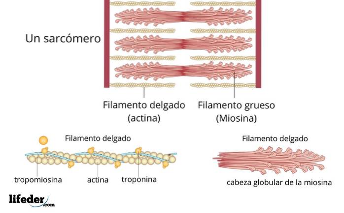
Myofibrils are long, fibrous structures and are made of two types of protein filaments that are stacked on top of each other..
Myosin is a thick fiber with a globular head, and actin is a thinner filament that interacts with myosin during the muscle contraction process..
A given myofibril contains approximately 10,000 sarcomeres, each of which is approximately 3 microns in length. Although each sarcomere is small, several aggregated sarcomeres span the length of the muscle fiber..
Each sarcomere consists of thick and thin bundles of the proteins mentioned above, which together are called myofilaments..
By enlarging a portion of the myofilaments, the molecules that compose them can be identified. The thick filaments are made of myosin, while the fine filaments are made of actin.
Actin and myosin are contractile proteins that cause muscle shortening when they interact with each other. In addition, the thin filaments contain other proteins with regulatory function called troponin and tropomyosin, which regulate the interaction between contractile proteins..
The main function of the sarcomere is to allow a muscle cell to contract. To do this, the sarcomere must be shortened in response to a nerve impulse..
The thick and thin filaments do not shorten, but instead slide around each other, causing the sarcomere to shorten while the filaments remain the same length. This process is known as the sliding filament model of muscle contraction..
The sliding of the filament generates muscle tension, which is undoubtedly the main contribution of the sarcomere. This action gives the muscles their physical strength..
A quick analogy for this is the way a long ladder can be extended or folded depending on our needs, without physically shortening its metal parts..
Fortunately, recent research offers a good idea of how this slip works. The sliding filament theory has been modified to include how myosin is able to pull on actin to shorten the length of the sarcomere..
In this theory, the globular head of myosin is located near actin in an area called the S1 region. This region is rich in hinged segments that can bend and thus facilitate contraction..
S1 bending may be the key to understanding how myosin is able to "walk" along actin filaments. This is achieved by cycles of myosin S1 fragment binding, contraction, and final release..
When myosin and actin join together, they form extensions called "cross bridges." These cross bridges can be formed and broken in the presence (or absence) of ATP, which is the energetic molecule that makes contraction possible..
When ATP binds to the actin filament, it moves it into a position that exposes its myosin-binding site. This allows the globular head of myosin to bind to this site to form the cross-bridge..
This binding causes the phosphate group of ATP to dissociate, and thus myosin begins its function. The myosin then goes into a lower energy state where the sarcomere can be shortened..
To break the cross-bridge and allow myosin to bind to actin again in the next cycle, the binding of another ATP molecule to myosin is necessary. That is, the ATP molecule is necessary for both contraction and relaxation..
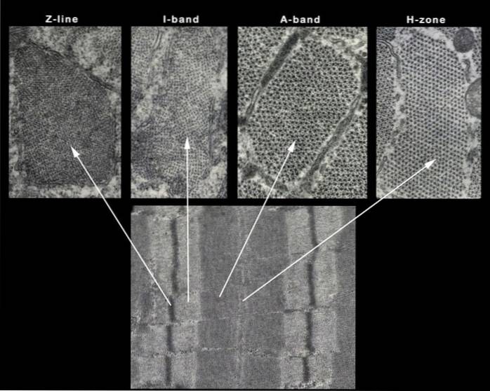
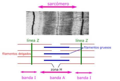
Histological sections of the muscle show the anatomical features of the sarcomeres. The thick filaments, composed of myosin, are visible and are represented as the A band of a sarcomere.
The thin filaments, made up of actin, bind to a protein in the Z disk (or Z line) called alpha-actinin, and are present throughout the length of the I band and a part of the A band.
The region where the thick and thin filaments overlap has a dense appearance, as there is little space between the filaments. This area where the thin and thick filaments overlap is very important for muscle contraction, as it is the site where the movement of the filament begins..
The thin filaments do not fully extend into the A bands, leaving a central region of the A band that contains only thick filaments. This central region of band A appears slightly lighter than the rest of band A, and is called zone H.
The center of the H zone has a vertical line called the M line, where accessory proteins hold the thick filaments together..
The main components of the histology of a sarcomere are summarized below:
Thick filament zone, composed of myosin proteins.
Central A-band zone, no overlapping actin proteins when muscle is relaxed.
Thin filament zone, composed of actin proteins (without myosin).
They are the boundaries between adjacent sarcomeres, formed by actin-binding proteins perpendicular to the sarcomere..
Central zone formed by accessory proteins. They are located in the center of the thick myosin filament, perpendicular to the sarcomere.
As mentioned earlier, contraction occurs when thick filaments slide along thin filaments in rapid succession to shorten myofibrils. However, a crucial distinction to remember is that the myofilaments themselves do not contract; it is the sliding action that gives them their power to shorten or lengthen.


Yet No Comments