
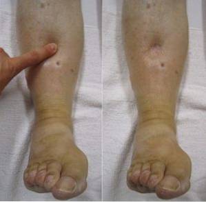
The godet sign or sign of the fovea is the sustained sinking of the skin when pressing on a point where there is a bony relief underneath, for example on the front of the leg. It is used in the physical examination of a patient with edema.
Edema is the accumulation of fluid in the subcutaneous cellular tissue and in the extracellular spaces of the body. This means that the amount of fluids in the tissue under the skin increases and the individual appears swollen..
There are several diseases that manifest with edema, either of a specific area or of the entire body. The most common cause of generalized edema is protein deficiency in the body, known as hypoproteinemia..
Swollen skin should be especially cared for as it can lead to changes such as dry skin, cracks and ulcers.
Article index
Godet's sign is always found in edematous patients and gives a clear diagnosis of edema and can even give information on how severe the inflammation is that this patient is presenting..
The way to perform the maneuver is by pressing the skin against a bony surface, for example on the front of the leg, for 5 seconds. If the skin is left with a cleft that takes a few seconds to return to its normal state, the sign is positive.
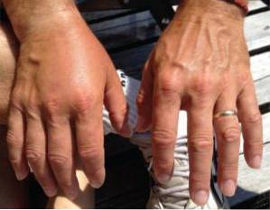
Edema is sought in the patient's declining points, that is, in those areas of the body that are closer to the ground. Thus, it is searched mainly in the hands, feet and legs, and in patients who are bedridden it is searched in the lower part of the back or in the ankles and back of the thighs..
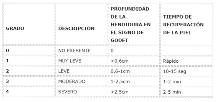
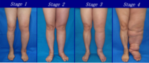
Edema is classified into four degrees according to the depth of the cleft that forms when the Godet sign is positive, and according to the time it takes for the skin to return to its normal state.
Edema is the accumulation of fluid outside the cells. It is formed through 4 pathophysiological mechanisms depending on the underlying disease of the patient.
Edema is considered one of the symptoms of a systemic condition and not a disease on its own.
The mechanisms of edema formation are as follows:
- Increased pressure of capillaries. Capillaries are small blood vessels that control pressure and the water system in the body. When there is an imbalance in this system, the kidneys retain fluid through a hormonal mechanism and this causes an increase in fluid in the tissues and edema..
- Decrease in the pressure exerted by the fluid outside the cell. This causes a pressure imbalance in which the cellular liquid can flow freely out because there is not enough force to stop it..
- Increased capillary permeability, which causes small blood vessels to increase their ability to pass fluid from the vascular space into the tissues. This mechanism of edema formation is common in infections.
- Lymphatic system obstruction. The lymph node and vessel system is a set of tubes that serve as a filter for some wastes in the body, such as large proteins or cells that are not used. When this system becomes obstructed, the fluid that normally circulates in your vessels (lymph) stagnates and begins to accumulate in the tissues below the obstruction..
The diagnosis of edema is clinical. The patient may have some symptoms before realizing that he has a swollen area of his body or that he has a general inflammation.
The symptoms described by the patient may be a sensation of weight, pressure, hot skin, changes in skin color and, in some cases, pain..
First of all, the questioning is important. It is necessary for the doctor to know the medical conditions of the patient, if there are any, since some diseases can have complications that end in edema or inflammation.
When the patient is questioned, it should be established when the inflammation started, what are the affected areas, if it occurs at a specific time and if it improves in any way. All this information guides the specialist in his diagnosis.
The physical examination should be a general examination that includes observation, measurement of the diameter of the arms and legs, and palpation of areas that appear inflamed. According to these characteristics, edema is classified into 4 grades: very mild, mild, moderate and severe..
Investigating the causes of edema can lead to the diagnosis of the disease that is causing it. It is important to order blood tests that include body proteins, urine tests, kidney function, heart function and finally X-ray and / or MRI images..
Edema due to problems of heart failure or disease occurs due to the imbalance of vascular pressures.
In general, the patient wakes up well and as the day goes by, they notice swelling, especially in the legs. Godet's sign is positive in these cases and is grade 2-3.
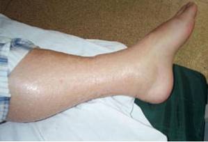
It occurs due to venous circulation problems and varicose veins. In these cases, the edema is associated with pain, tenderness and changes in the skin of the legs. In advanced cases, the skin may be broken and form ulcers that secrete yellowish fluid..
Godet's sign is easily evidenced in grade 2-3 soft edema, with pain on pressure..
Lymphedema occurs when there is obstruction of the lymphatic system. In these cases, the edema is seen below the obstruction, for example if the patient has a tumor in the armpit, the arm on that side will begin to swell.
Lymphedema gets worse and worse if the blockage is not removed. The main causes are tumors, benign or malignant, and surgeries in which the lymph nodes are removed as in some types of breast cancer surgery.
The most extreme case is that of infection by the parasite called filaria. This parasite obstructs the lymphatic vessels of the skin causing a syndrome called lymphatic filariasis or elephantiasis..
All cases of lymphedema present with obvious Godet's sign, with a grade 4 deep cleft, and the edema is usually hard..
There are some medications that when used for long periods of time can lead to true states of inflammation.
It occurs with some antidepressants and anti-inflammatories. Edema improves by eliminating the administration of these drugs. Godet's sign may be weak, grade 1-2, but is present.
The decrease in proteins in the plasma fluid, which is part of the blood, causes an imbalance of pressures and that cellular fluid begins to come out and even begins to be excreted in the urine. In these cases, the nutritional condition of the patient should be evaluated.
The type of inflammation observed in cases of hypoproteinemia is generalized edema that does not improve until the proteins that are decreased are replaced.
Godet's sign is always found in these cases, the edema is soft and depending on the time of the disease the cleft formed in the skin can be deep.

Yet No Comments