
The homans sign is the pain response in the lower limbs to a maneuver performed by the doctor in the physical examination of a patient in whom vascular, specifically venous insufficiency is suspected.
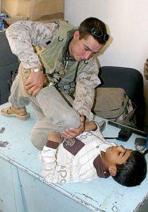
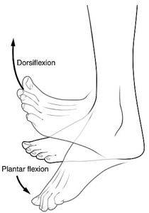
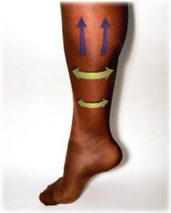
The maneuver consists of passively moving the patient's foot from the ankle joint, so as to achieve dorsiflexion of the ankle. This movement should be done quickly and firmly, but carefully.
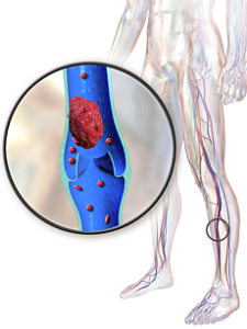
The sign is considered positive when the person manifests flexion pain, and it is one of the tests performed in patients with deep vein thrombosis (DVT). This medical condition is a condition in which a blood clot forms and blocks the deep veins. It is frequently evidenced in the veins of the lower limbs, below the knees, but can occur in any part of the body.
The causes that trigger a deep vein thrombosis are very varied and, although genetic disorders can be observed that increase the risk of suffering from this condition, they are generally acquired.
Article index
Also known as the sign of dorsiflexion, it is a physical test that consists of triggering pain on passive flexion of the lower limb, in a patient in whom DVT is suspected. It is a semiological sign that is looked for in the physical examination.
It was described in 1944 by Dr. John Homans (1877-1954), an American surgeon at Massachusetts General Hospital in Boston, who dedicated his surgical career to the study of vascular diseases..
To look for the sign, a maneuver must be performed in which the doctor first tells the patient to lie on his back. In this position, the same examiner raises the patient's leg, leaving the knee slightly flexed, and proceeds to mobilize the ankle joint until the foot is flexed..
The movement should be fast and firm but gentle enough so that it does not cause trauma or injury..
The sign is considered positive if the patient reports pain in the calf, or behind the knee, when the foot is dorsiflexed..
This painful response occurs because the calf muscles contract and press on the deep tibial vein, which is swollen and weak with DVT..
The Homans sign is a resource to the physical examination that is taken into account in case the examiner suspects DVT. However, it is a test that is not specific, that is, it can occur in other clinical conditions, and it can be negative in patients with the disease..
Currently the diagnosis of DVT is made through non-invasive imaging methods such as venous ecosonogram and vascular magnetic resonance..
For this reason, a diagnosis should not be established, nor should a medical therapy be indicated, just because of the positive finding of this sign..
Deep vein thrombosis (DVT) is a pathology characterized by the abnormal formation of a clot that obstructs the blood flow of the deep veins of the body..
This condition must be diagnosed in time to be able to administer the patient the opportune treatment and thus avoid complications, which can be fatal..
DVT can occur in any of the veins that run deep in the body, however, the most common is that it occurs in the lower limbs specifically below the knees.
The leading cause of death from DVT is pulmonary embolism, condition in which the clot formed in the veins travels to the lung obstructing the circulation of that organ.
The disease has multiple causes, both environmental and genetic. One of the most frequent is the decrease in venous blood flow due to immobility.
The person who is immobilized, either due to injury to the lower limbs, fractures for example, or due to any condition that prevents ambulation, such as chronic diseases that lead to fatigue, regardless of age, should receive prophylactic or preventive therapy to TVP.
Likewise, healthy patients from a cardiovascular point of view who must undergo surgeries lasting more than 3 hours, or who must stay on a flight for more than 4 hours, should take preventive measures.
Some of the measures are the subcutaneous injection of anticoagulants and the use of anti-embolic stockings, which are special stockings that put continuous pressure on the leg to maintain blood flow..
DVT is suspected in those patients with a history or significant diseases that are at risk for thrombosis and present with pain and redness in one or both lower limbs..
The diagnostic approach begins with the questioning of the patient. It should be evaluated if the patient has been exposed to those elements that are considered risk factors. For example, a person with pain in a lower limb after an interoceanic trip.
Obesity is a risk factor for DVT, so it is important to know the body mass index (BMI) of the patient, whose calculation is made knowing the weight and height (BMI = weight in Kg ÷ height in meterstwo). Also pregnant patients; with voluminous abdominal tumors, and those lesions that limit mobility, are prone to present the condition.
Once this information is available, a physical examination is carried out, which can provide important data to reach the final diagnosis. The veins of the lower limbs are usually the most affected in an episode of DVT, therefore during the physical evaluation emphasis is placed on this area, especially in the limb that is believed to be affected.
The best way to evaluate an even member is to compare it to the other. For example, to know with certainty if one leg is swollen, the circumference of both can be measured and see if they coincide. Likewise, skin color and temperature are parameters that are compared to evaluate the condition of the limb in which DVT is suspected..
In addition to this, there are physical maneuvers that the doctor can practice to trigger pain. From these, the semiological signs for DVT can be highlighted..
These signs include Olow's, Lowenberg's, and Homans's among others. All focus on trying to produce lower limb pain with passive leg mobilization maneuvers.


Yet No Comments