
The McBurney sign is the pain caused by the doctor in the abdominal physical examination at the McBurney point, one of the points explored in patients with appendicitis. It is one of the answers that one tries to obtain in order to reach a proper diagnosis in a patient with abdominal pain..
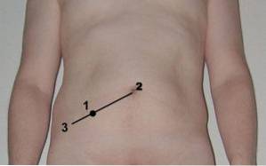
To observe the McBurney sign, the doctor must identify the point of maximum abdominal pain in appendicitis, or McBurney point. This point is located at the junction between the internal two thirds with the external third of an imaginary line drawn between the umbilicus and the right antero-superior iliac spine..
In the patient with acute appendicitis, this area may be tender. But sometimes this is not the case, so pressure exerted on the McBurney point is used to generate pain due to irritation of the layer that covers the abdomen (peritoneum).
Although the McBurney sign is not specific for acute appendicitis, it is a reliable indicator that there is a serious inflammatory process in the abdomen and that it needs to be treated as immediately as possible..
Article index
The McBurney sign is one of the most important in the abdominal physical examination in the patient with acute pain. It was described in 1889 by Dr. Charles McBurney, a surgeon and professor at Rossevelt Hospital in New York. In the article in which he explains the sign, he also describes the location of the McBurney point..
In his work Experience with early operative interference in cases of vermiform appendix disease (1889) Dr. McBurney stated:
"The place of greatest pain, determined by the pressure of a single finger, has been very exact between one third and two thirds from the anterior superior iliac spine, drawing a straight line to the navel "
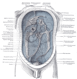
Peritonitis refers to inflammation of the deep layer that lines the abdominal cavity called the peritoneum. It occurs due to acute inflammation of an intra-abdominal organ.
The peritoneum is a semi-permeable layer that lines the abdomen. It contains only enough cellular liquid for the two layers that make it up to slide off each other. Its balance is altered when bacteria from a contaminated intra-abdominal organ pass into the cavity or when an organ is perforated.
Faced with contamination, the peritoneum produces more fluid than normal and a true inflammatory process begins that manifests itself with acute abdominal pain. The thoracic nerves are those that innervate this area and those that are responsible for sending impulses that manifest as pain.
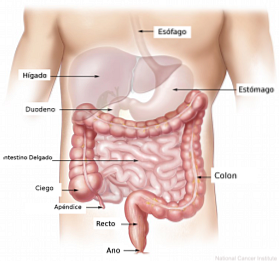
The most common pathologies associated with peritonitis are acute appendicitis, inflammation of the cecal appendix and acute cholecystitis or inflammation of the gallbladder..
Depending on the organ that is causing the peritonitis, different signs may be found in the patient on the medical physical examination, some more accurate than others..
The physical examination in peritonitis is usually nonspecific, since the nerve fibers of the organs do not localize pain well. Therefore, the patient may have a pelvic process and locate pain throughout the abdomen without being able to specify a site. This is called referred pain..
Inflammation of the cecal appendix is the most common inflammatory process in the abdomen and the leading cause of peritonitis. It is an acute process that is fully established between 6 and 8 hours and that can put the patient's life at risk.
The diagnosis of acute appendicitis is basically clinical. This means that the doctor must rely on the questioning and pay special attention to the physical examination and laboratory tests of the patient..
Within the physical examination of acute appendicitis, different ways of evaluating appendicular pain have been described. Most of the tests used are named after the doctor who described them..
Thus, we find the Rovsing sign, the Owen sign and the McBurney sign, among many others. The tests consist of trying to locate the pain in the right iliac fossa, which is the site where the cecal appendix is located..
To reach the diagnosis of appendicitis it is important to know that it is an acute process that can take up to 8 hours to fully establish.
The triad of abdominal pain that migrates from the navel to the right iliac fossa, the lack of appetite and altered blood tests, can guide the doctor to the definitive diagnosis.
Identifying a water appendicitis is of the utmost importance since it is a disease that contaminates the abdominal cavity. Over the hours this contamination can reach the blood and can be lethal if it is not treated in time. Treatment is surgical.
Abdominal palpation is difficult and requires experience to be able to verify or rule out a diagnosis.
The doctor must know well the anatomy of the intra-abdominal organs and their anatomical projection on the patient's abdomen, as well as the pathophysiological process of the most common diseases of the abdomen in order to reach a diagnosis.
In the case of appendicitis, more than twenty maneuvers have been described to show appendicular pain. Although none are totally specific to appendicitis, it is important to know them in order to be able to perform them correctly and reach the diagnosis.
Acute appendicitis is a surgical emergency. When diagnosed, the patient must undergo surgery to remove this organ..
The most commonly used incision for the surgical approach to this pathology was also described by Charles McBurney. It involves incising the skin of the abdomen with an oblique wound, over the McBurney point.
It is assumed that because the McBurney point is located where the cecal appendix is in most patients, when the McBurney incision is made, there is complete and perfect access to remove it..
Although this is the most popular incision, other surgical techniques have been described with equal exposure and better aesthetic results..
Currently, in most cases, it is preferred to carry out the removal of the appendix through laparoscopic surgery. In this type of surgery, 4 small incisions are made through which special instruments are introduced to complete the procedure..


Yet No Comments