
The Rovsing sign it is a sign present in the abdominal physical examination of a person with peritoneal irritation, especially oriented towards an inflammatory process of the cecal appendix. This sign is explored in patients with abdominal pain and should be taken into account for the diagnostic approach.
Described in 1907 by Dr. Niels Thorkild Rovsing, the sign consists of putting pressure on the left iliac fossa, which will generate an increase in pressure in the right colon causing pain in the cecal appendix, which is located on that side..
Although Rovsing's sign is not specific for appendicitis, it does translate as the sign produced by a disease of the right iliac fossa. In the case of women, these processes can be a ruptured or bleeding ovarian cyst or a ureter stone, among others..
Article index
Dr. Niels Thorkild Rovsing was a remarkable abdominal surgeon. Among his professional milestones, he came to describe surgical techniques and signs of the physical examination that he commonly observed in his patients..
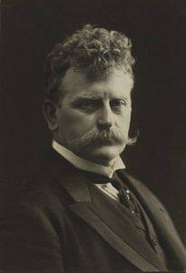
In 1907 he described a sign of peritoneal irritation that many patients diagnosed with acute appendicitis presented. This was reflected in his work Indirect evocation of typical McBurney point pain. A contribution to the diagnosis of appendicitis and typhillitis.
The idea was to press the descending colon by insufflating the right colon and, in this way, stimulate the right side of the colon causing pain..
The maneuver has been used widely, so it is common to hear the eponymous among surgeons at the time of an abdominal physical examination..
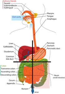
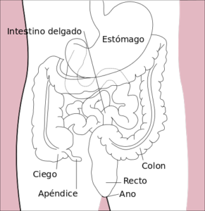
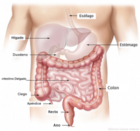
The vermiform appendix or cecal appendix, is an elongated organ that is connected to the first portion of the large intestine, called the cecum.
It is a cylindrical structure, without an exit hole. It is believed that it is a vestigial organ, that is, that evolutionarily it has been atrophying until this small intestinal remnant remains. The appendix is located in the right iliac fossa, which is the lower right area of the abdomen.
Its functions are not fully known, although it is believed that in the past it was an important organ in the digestion of some vegetables. The functions that have been attributed to it in modern medicine include the capacity of lymphatic drainage and maintaining the intestinal flora of the colon..
Appendicitis is the inflammatory process that occurs in the appendix for causes as diverse as, for example, the presence of a fecalite. This is a small compact amount of feces that obstructs the appendicular orifice or external bacterial processes such as tuberculosis.
Inflammation of the appendix is the most common cause of appendicular pathologies, although malignant processes such as carcinoid tumors can also occur.
The treatment of appendicitis is surgery, and it must be treated at the time it is diagnosed. Surgery can be performed by the conventional route in an open way, or by laparoscopy..
The diagnosis of appendicitis is always clinical. This means that there are no special diagnostic tests that provide the doctor with a more accurate diagnosis than that provided by the abdominal physical examination and a blood test..
The doctor must question the patient in order to rule out some diagnoses. For example, in patients between 13 and 25 years old, with diffuse abdominal pain that is located in the right iliac fossa and presents loss of appetite and vomiting, there is a high suspicion of acute appendicitis.
The blood test shows the typical values of a bacterial infection, elevated white blood cells with a large percentage of neutrophils. White blood cells are the blood cells that indicate infection, while neutrophils are specialized white blood cells, especially active in bacterial infectious processes..
Diagnostic confirmation is made through physical examination. What is sought is to perform the so-called appendicular maneuvers and highlight the pain in the right iliac fossa.
When the doctor suspects the diagnosis of acute appendicitis, they go to the physical examination to reveal the pain in the right iliac fossa that causes the appendicitis.
There are many maneuvers that can be performed, the most common being the McBurney sign, the rebound sign, and the Rovsing sign itself..
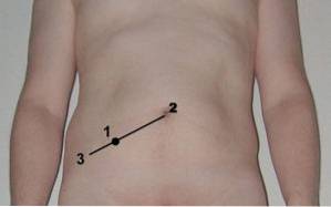
The McBurney sign is perhaps the most used and consists of causing pain by pressing on the McBurney point, which is the point where the appendix is located topographically.
To locate the McBurney point, an imaginary line is drawn between the navel and the iliac crest and the union between the internal two thirds with the external third is sought.
The rebound sign is achieved by pressing on any part of the abdomen and releasing the hand with a sudden movement. This causes the two layers of peritoneum to bounce off each other, causing pain. It is not a specific sign of appendicitis.
In the specific case of the Rovsing sign, Dr. Rovsing described it in 1907 as the pressure of the left colon to fill the right colon with air and thus cause pain.
In other words, the left colon must be compressed, trying to transfer air to at least the transverse colon. With this, try to fill the cecum with air and that compression caused by the air causes pain in the irritated appendix.
Currently, the maneuver to find the Rovsing sign has been changed and for practicality only seeks to provoke reflex pain. This type of pain occurs because the neurological connections of the peritoneum, the layer that lines the abdominal cavity, are not as precise to capture the pain.
This means that if the inflammatory process is on the right side, when touching any point of the abdomen the patient will feel the pain on the right side..
However in Dr. Rovsing's original work the maneuver is clearly described. This begins by placing the left hand on the left side of the patient's lower abdomen, where the colon is supposed to be, and the right hand on top. With the right hand, the abdomen is pressed and an upward movement begins throughout the left abdomen..
The idea is to move the air that is inside the left colon to the right colon. This increases the pressure on that side and this will cause pain in the diseased appendix..
Rovsing's sign may be positive in other processes that cause inflammation in the right iliac fossa, such as inflammation of the ascending colon and inflammatory processes of the ovaries..


Yet No Comments