
The neuronal synapse It consists of the union of the terminal buttons of two neurons in order to transmit information. In this connection, a neuron sends the message, while one part of the other receives it..
Thus, communication usually occurs in one direction: from the terminal button of a neuron or cell to the membrane of the other cell, although it is true that there are some exceptions. A single neuron can receive information from hundreds of neurons.
Each single neuron receives information from the terminal buttons of other nerve cells, and in turn the terminal buttons of the latter make synapses with other neurons.
Article index
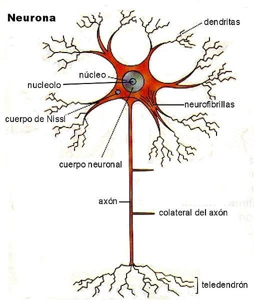
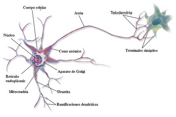
The terminal button is defined as a small thickening at the end of an axon, which sends information to the synapse. Whereas, an axon is a kind of elongated and thin "wire" that carries messages from the nucleus of the neuron to its terminal button..
The terminal buttons of nerve cells can synapse with the membrane of the soma or dendrites.
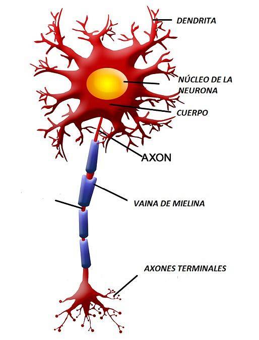
The soma or cell body contains the nucleus of the neuron; It has mechanisms that allow the maintenance of the cell. Instead, dendrites are tree-like branches of the neuron that start from the soma..
When an action potential travels through the axon of a neuron, the terminal buttons release chemicals. These substances can have excitatory or inhibitory effects on the neurons with which they connect. At the end of the whole process, the effects of these synapses give rise to our behavior.
An action potential is the product of communication processes within a neuron. In it there are a set of alterations in the axon membrane that cause the release of chemicals or neurotransmitters.
Neurons exchange neurotransmitters at their synapses as a way to send information to each other.
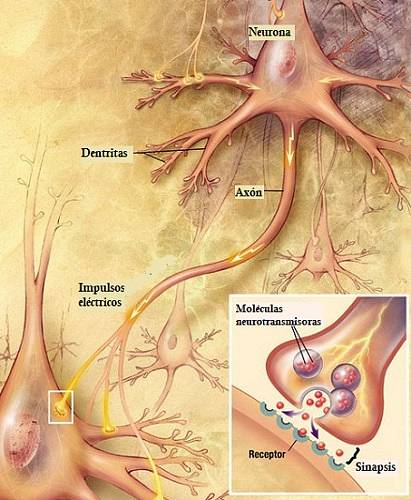
Neurons communicate through synapses, and messages are transmitted by releasing neurotransmitters. These chemicals diffuse into the liquid space between the terminal buttons and the membranes that establish synapses..
The neuron that releases neurotransmitters through its terminal button is called a presynaptic neuron. While the one that receives the information is the postsynaptic neuron.
When the latter captures neurotransmitters, the so-called synaptic potentials are produced. That is, they are alterations in the membrane potential of the postsynaptic neuron..
To communicate, cells must secrete chemicals (neurotransmitters) that are detected by specialized receptors. These receptors consist of specialized protein molecules.
These phenomena are simply differentiated by the distance between the neuron that releases the substance and the receptors that capture it..
Thus, neurotransmitters are released by the terminal buttons of the presynaptic neuron and are detected through receptors located on the membrane of the postsynaptic neuron. Both neurons must be located within a short distance for this transmission to occur..
However, contrary to popular belief, neurons that make chemical synapses do not physically join. In fact, between them there is a space known as the synaptic space or synaptic cleft..
This space appears to vary from synapse to synapse, but is generally about 20 nanometers wide. There is a network of filaments in the synaptic cleft that keeps pre and postsynaptic neurons aligned..
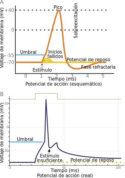
For an exchange of information to occur between two neurons or neuronal synapses, an action potential must first occur.
This phenomenon occurs in the neuron that sends the signals. The membrane of this cell has an electrical charge. In reality, the membranes of all cells in our body are electrically charged, but only axons can provoke action potentials.
The difference between the electrical potential inside the neuron and outside is called the membrane potential..
These electrical changes between the inside and outside of the neuron are mediated by existing concentrations of ions, such as sodium and potassium..
When there is a very rapid reversal of the membrane potential, an action potential occurs. It consists of a brief electrical impulse, which the axon conducts from the soma or nucleus of the neuron to the terminal buttons.
It should be added that the membrane potential must exceed a certain excitation threshold for the action potential to occur. This electrical impulse is translated into chemical signals that are released through the terminal button..
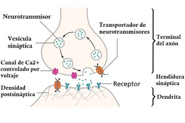
Neurons contain sacs called synaptic vesicles, which can be large or small. All terminal buttons have small vesicles that carry neurotransmitter molecules inside..
Vesicles are produced in a mechanism located in the soma called the Golgi apparatus. They are then transported close to the terminal button. However, they can also be produced on the terminal button with "recycled" material..
When an action potential is sent along the axon, depolarization (excitation) of the presynaptic cell occurs. As a consequence, the calcium channels of the neuron open allowing calcium ions to enter it..

These ions bind to molecules on the membranes of the synaptic vesicles that are on the terminal button. Said membrane breaks, fusing with the membrane of the terminal button. This produces the release of the neurotransmitter into the synaptic space..
The cell's cytoplasm captures the remaining pieces of membrane and carries them to the cisterns. There they are recycled, creating new synaptic vesicles with them..

The postsynaptic neuron has receptors that capture substances that are in the synaptic space. These are known as postsynaptic receptors, and when activated, they cause ion channels to open..
When these channels open, certain substances enter the neuron, causing a postsynaptic potential. This can have excitatory or inhibitory effects on the cell depending on the type of ion channel that has been opened.
Normally, excitatory postsynaptic potentials occur when sodium enters the nerve cell. While the inhibitors are produced by the exit of potassium or the entry of chlorine.
The entry of calcium into the neuron causes excitatory postsynaptic potentials, although it also activates specialized enzymes that produce physiological changes in this cell. For example, it triggers the displacement of synaptic vesicles and the release of neurotransmitters.
It also facilitates structural changes in the neuron after learning..
Postsynaptic potentials are usually very brief and terminate through special mechanisms.
One of them is the inactivation of acetylcholine by an enzyme called acetylcholinesterase. Neurotransmitter molecules are removed from the synaptic space by reuptake or reabsorb by transporters that are in the presynaptic membrane.
Thus, both presynaptic and postsynaptic neurons have receptors that capture the presence of chemicals around them..
There are presynaptic receptors called autoreceptors that control the amount of neurotransmitter released or synthesized by the neuron.

In them an electrical neurotransmission takes place. The two neurons are physically connected through protein structures known as gap junctions..
These structures allow changes in the electrical properties of one neuron to directly influence the other and vice versa. In this way, the two neurons would act as if they were one..
Chemical neurotransmission occurs at chemical synapses. Pre and postsynaptic neurons are separated by the synaptic space. An action potential in the presynaptic neuron would cause the release of neurotransmitters.
These reach the synaptic cleft, being available to exert their effects on postsynaptic neurons.
An example of an excitatory neuronal synapse would be the withdrawal reflex when we burn out. A sensory neuron would detect the hot object, since it would stimulate its dendrites.
This neuron would send messages through its axon to its terminal buttons, located in the spinal cord. The terminal buttons of the sensory neuron would release chemicals known as neurotransmitters that would excite the neuron with which it synapt. Specifically, to an interneuron (the one that mediates between sensory and motor neurons).
This would cause the interneuron to send information along its axon. In turn, the terminal buttons of the interneuron secrete neurotransmitters that excite the motor neuron..
This type of neuron would send messages along its axon, which attaches to a nerve to reach the target muscle. Once neurotransmitters are released by the terminal buttons of the motor neuron, the muscle cells contract to move away from the hot object..
This type of synapse is somewhat more complicated. It would be given in the following example: imagine that you take a very hot tray out of the oven. You wear mittens to avoid burning yourself, however, they are somewhat thin and the heat begins to overcome them. Instead of throwing the tray on the ground, you try to withstand the heat a bit until you put it on a surface.
The withdrawal reaction of our body to a painful stimulus would have made us let go of the object, even so, we have controlled this impulse. How is this phenomenon produced?
The heat coming from the tray is perceived, increasing the activity of the excitatory synapses on the motor neurons (as explained in the previous section). However, this excitement is counteracted by inhibition that comes from another structure: our brain..
It sends information indicating that if we drop the tray, it could be a total disaster. Therefore, messages are sent to the spinal cord that prevent the withdrawal reflex..
To do this, an axon from a brain neuron reaches the spinal cord, where its terminal buttons synapse with an inhibitory interneuron. It secretes an inhibitory neurotransmitter that reduces motor neuron activity, blocking the withdrawal reflex..
Importantly, these are just examples. The processes are really more complex (especially the inhibitory ones), with thousands of neurons involved in them.
- Axodendritic synapses: in this type, the terminal button connects to the surface of a dendrite. Or, with dendritic spines, which are small protrusions located on the dendrites in some types of neurons.
- Axosomatic synapses: in these, the terminal button synapt with the soma or nucleus of the neuron.
- Axoaxonic synapses: the terminal button of the presynaptic cell connects with the axon of the postsynaptic cell. This type of synapse works differently from the other two. Its function is to reduce or boost the amount of neurotransmitter that is being released by the terminal button. Thus, it promotes or inhibits the activity of the presynaptic neuron.
Dendrodendritic synapses have also been found, but their exact role in neuronal communication is not currently known..
During neuronal communication, not only neurotransmitters such as serotonin, acetylcholine, dopamine, norepinephrine, etc. are released. Other chemicals such as neuromodulators can also be released.
These are so named because they modulate the activity of many neurons in a certain area of the brain. They secrete in greater quantity and travel longer distances, spreading more widely than neurotransmitters.
Another type of substance is hormones. These are released by cells of the endocrine glands, which are located in different parts of the body such as the stomach, intestines, kidneys and brain..
Hormones are released into the extracellular fluid (outside the cells), and are subsequently taken up by capillaries. They are then distributed throughout the body through the bloodstream. These substances can bind to neurons that have special receptors to take them up..
Thus, hormones can affect behavior, altering the activity of the neurons that receive them. For example, testosterone appears to increase aggressiveness in most mammals..


Yet No Comments