The portal system It is a type of specialized circulation that connects two anatomical structures in order to transport specific substances beyond nutrients and oxygen. It is a very specialized type of circulation present in very specific regions where it fulfills a well-defined function, in fact in humans there are only two portal systems: the hepatic and the hypothalamic-pituitary.
The main characteristic of portal circulation is that it begins and ends in venous capillaries. It differs from the general systemic circulation in that the latter usually begins in arterial components that progressively decrease in caliber; once the arterial capillary level is reached, the venous segment of the circuit begins to be built, from the venous capillaries, passing through the venules until reaching the veins.
For their part, portal systems start as venous capillaries that emerge from a structure, join together to form a vein, which will again divide into hundreds of venous capillaries at the other end of the system..
Another particular characteristic of portal circulation is that it is an exclusively venous system, that is, there are no arteries involved in the formation of the system..
Article index
In general, the systemic circulation has two components, an arterial one that carries oxygen and nutrients to the tissues, and a venous one that collects waste that will be eliminated in the liver and kidney, also carrying non-oxygenated blood to the lung where the exchange will take place. carbon dioxide for oxygen.
However, when specific substances other than oxygen and nutrients need to be transported between two distant anatomical regions, it is necessary for the body to "channel" them into a specific and direct transport system..
In this way, the substances to be transported do not spread throughout the body through the general circulation, but rather go from point A to point B in an expeditious manner..
Since it is a very specialized type of circulation, portal systems are not common in humans, in fact there are only two:
- Hepatic portal system
- Hypothalamic-pituitary portal system
According to its anatomical location, the portal circulation is intended for the transport of specific substances between two target points, as indicated below:
Its objective is to transport the macronutrients absorbed in the intestine to the liver, where they will be converted into products that can be used by the rest of the organs and systems..
It constitutes a direct blood connection between two areas of the central nervous system that communicate and regulate each other between chemical mediators.
The inducing hormones released in the hypothalamus reach the pituitary directly through the hypothalamic-pituitary portal circulation. Once there, they induce the production of specific hormones in the anterior pituitary, which are released into the circulation..
Through systemic circulation these hormones reach the hypothalamus where they inhibit the production of the inducing hormone (negative feedback system).
The common denominator of portal circulation is the fact of being venous and having its beginning and end in a capillary network, however, depending on its location, the anatomy of each portal system varies ostensibly.
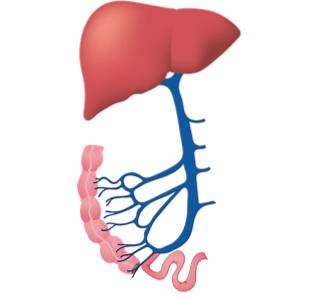
The capillaries that give rise to it are found in the submucosa of the small intestine where the nutrients absorbed in the intestine reach the circulation.
These capillaries join together to give rise to venules in the thickness of the intestinal wall, which in turn converge to form a complex venous network in the intestinal meso.
All these veins converge to form the superior and inferior mesenteric veins, which in their course unite, also receiving the splenic vein and sometimes the left gastric vein, giving rise to the portal vein..
The portal vein runs in direct relation to the posterior aspect of the pancreas, then ascends parallel to the bile duct and the hepatic artery where they divide into left and right lobar branches..
The lobar branches are subdivided into segmental branches to finally give their terminal branches at the level of the hepatic sinusoids, where finally the blood can release the nutrients towards the hepatocytes to be processed.
The hepatic portal system is large and complex, extending for a considerable distance into the abdominal cavity and transporting vast amounts of nutrients..
Unlike its hepatic counterpart, the hypothalamic-pituitary portal is a very short and localized system, in fact the hypothalamic-pituitary vein is less than 1 cm in length.
Despite its importance, the anatomical details of this system are not as fully understood as those of the hepatic portal. However, broadly speaking, it can be said that the capillaries that give rise to this system are found in the thickness of the hypothalamus, where they receive the inducing hormones that must be transported to the pituitary..
The different capillaries that make up this wide network join together to give rise to the hypothalamic-pituitary portal vein, which runs parallel to the pituitary pedicle..
Once it reaches the anterior lobe of the pituitary, this vein divides again into several thousand venous capillaries that carry the inducing hormones directly to the effector cells located in the adenohypophysis..
The best known disease that affects the portal system is portal hypertension, which occurs in the hepatic portal system.
Portal hypertension occurs when there is obstruction of the outlet capillaries at the hepatic end of the system. The obstruction can be before the sinusoidal capillaries, in the capillaries themselves or beyond them, in the hepatic veins.
When the obstruction is before the sinusoidal capillaries, portal hypertension is classified as presinusoidal, the main cause being schistosomiasis (previously known as bilharzia)..
In this disease, the adult forms of the schistosoma (a flatworm worm) reach the mesenteric venules, settling in them to fulfill their life cycle.
The presence of these small worms that do not exceed 10 mm in length obstructs the capillary plexuses, thus increasing the pressure between the origin of the portal system and the point of obstruction.
In cases where the problem is located in the hepatic sinusoidal capillary (sinusoidal portal hypertension), the reason is usually fibrosis associated with cirrhosis (which in turn induces sclerosis of the vascular elements) or liver cancer with associated destruction of the anatomical structures.
Finally, when the obstruction is located beyond the terminal portal capillaries, in the suprahepatic veins or the inferior cava, it is referred to as postsinusoidal portal hypertension, the most common cause being thrombosis of the suprahepatic veins and Budd-Chiari syndrome..
Portal hypertension is clinically characterized by the presence of ascites (free fluid in the abdominal cavity) associated with the development of a venous network collateral to the portal system..
This venous network is found in the rectum (hemorrhoidal plexuses), the esophagus (cardio-esophageal veins), and the abdominal wall (epigastric veins)..
Depending on the type of hypertension, other symptoms may be associated, the most frequent being jaundice (yellowing of the skin and mucous membranes) in cases of sinusoidal portal hypertension and edema in the lower limbs in cases of postsinusoidal portal hypertension..
Treatment of portal hypertension should be aimed at correcting the cause whenever possible; When this cannot be carried out, palliative treatments should be chosen aimed at reducing the pressure in the system..
For this, there are various surgical techniques which share one characteristic in common: the creation of a porto-systemic shunt to relieve pressure on the portal system..


Yet No Comments