
The Apert syndrome or acrocephalosyndactyly type I (ACS1) is a pathology of genetic origin that is characterized by the presence of different alterations and malformations in the skull, face and extremities.
At a clinical level, Apert syndrome is characterized by the presence or development of a pointed or elongated skull, sunken facial area with an alteration in the projection of the teeth, fusion and closure of the finger bones and joints, mental retardation variable, language disturbances, etc..
Although this pathology can be hereditary, in most cases Apert syndrome occurs without the presence of a family history, essentially due to a de novo mutation during the gestation phase..
The genetic mechanisms that cause Apert syndrome are not exactly known. Currently, various genetic alterations that are capable of producing this pathology have been identified, essentially related to mutations in the FGFR2 gene.
On the other hand, the diagnosis of Apert syndrome usually begins with clinical suspicion in the prenatal period after the identification of abnormalities in routine ultrasound scans and is confirmed by conducting a genetic study.
Regarding treatment, there is no type of curative intervention for Apert syndrome. However, throughout the history of this pathology, various specific interventions have been designed, which usually include neurosurgery, craniofacial surgery, maxillofacial surgery, pharmacological treatment, physical therapy, psychological and neuropsychological intervention, among others..
Article index
Apert syndrome is a genetic pathology characterized by the presence of different skeletal malformations at the cranial, facial and / or limb level.
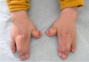
The essential alteration of Apert syndrome is constituted by a premature or early closure of the cranial fissures, which causes an abnormal growth of the rest of the structures of the face and skull. In addition to these, malformations may also appear in the upper and lower extremities, such as the fusion of the fingers and toes..
On the other hand, the cognitive abilities of people suffering from Apert syndrome may also be affected, with a variable severity from mild to moderate.
Although Baumgartner (1842) and Wheaton (1894) made the first mentions about this medical condition, it was not until 1906, when the French medical specialist Eugene Apert, accurately described this syndrome and published the first clinical report..
In his publication, Eugene Apert, describes a set of new cases of patients affected by a well-defined malformative pattern and characterized by the characteristic signs and symptoms of this pathology..
Thus, it was not until 1995 that the etiological genetic factors of Apert syndrome were identified. Specifically, Wilkie et al. Described the presence of two mutations in the FGFR2 gene in around 40 affected patients..
In addition, Apert syndrome is a medical condition that is classified within the diseases or pathologies characterized by presenting craniosynostosis (premature closure of cranial sutures).
Other pathologies belonging to this group are Pfeiffer syndrome, Crouzon syndrome, Saethre-Chotzcen syndrome and Carpenter syndrome..
Apert syndrome is considered a rare or infrequent pathology, that is, it has a prevalence of less than one case per 15,000 inhabitants of the general population.
Specifically, Apert syndrome occurs around one person for every 160,000-200,000 births and, in addition, there is a 50% probability of transmitting this pathology at the hereditary level.
Furthermore, in terms of gender distribution, a higher prevalence in men or women has not been identified, nor has it been associated with particular ethnic groups or geographic locations..
Currently, and given that Apert syndrome was identified in approximately 1984, in clinical reports and in the medical literature that have published more than 300 cases of this pathology.
The clinical manifestations of Apert syndrome usually include a malformation or incomplete development of the cranial structure, an atypical phenotype or facial pattern, and skeletal changes in the extremities..
In the case of Apert syndrome, the central involvement is related to the formation and closure of the bone structure of the skull. During embryonic development, a process called creneosynostosis occurs, characterized by premature closure of the cranial sutures.
Cranial fissures or sutures are a type of fibrous tissue bands that have the fundamental objective of connecting the bones that make up the skull (frontal, occipital, parietal and temporal).
During the gestation phase and the early postnatal period, the bone structure that makes up the skull is held together thanks to these fibrous and elastic tissues.
Normally, the cranial bones do not fuse until around 12 to 18 months. The presence of soft spots or spaces between the cranial bones is part of normal child development.
Therefore, during the entire childhood stage, these sutures or flexible regions allow the brain to grow in an accelerated way and, in addition, protect it from impacts.
Thus, in Apert syndrome, premature closure of these cranial sutures and cranial bones makes normal development of cranial and brain growth impossible..
Consequently, the most common signs and symptoms of Apert syndrome may include:
These types of alterations mainly affect the upper and lower extremities, usually the fusion and development of the fingers..
In addition to these, it is also possible to observe other clinical findings at the musculoskeletal level, shortening of various bones (radius, humerus, femur), hypoplasia of the scapula or pelvis, fusion of cervical vertebrae.
As a consequence, many affected will present reduced joint mobility and, therefore, may develop various difficulties for the acquisition of gross and fine motor skills.
These types of anomalies are very heterogeneous and variable among affected individuals, however, some of the most common have been identified:
The etiological alteration of this pathology can lead to the development of lesions or secondary pathologies at a morphological and structural level in various areas of the body, some of them include:
Despite the fact that in many cases it is possible to observe the presence of a general alteration of cognitive functions and intellectual level, mental retardation is not unequivocally present in all cases of Apert syndrome..
In addition, in cases where there is an impairment of the intellectual level, this can be variable, on a scale from mild to moderate.
On the other hand, in the linguistic area, the development of various deficits is frequent, mainly related to the articulation of sounds as a result of mandibular and oral malformations..
Apert syndrome is due to the presence of a specific mutation in the FGFR2 gene. Experimental studies have indicated that this gene is responsible for the production of a protein, called fibroblast growth factor receptor 2.
Among the functions of this factor, the sending of different chemical signals to immature cells is described to cause their transformation and differentiation into bone cells during the fetal or prenatal phase of development..
Therefore, the presence of mutations in the FGFR2 gene alters the functioning of this protein and, therefore, can cause the early fusion of the bones of the skull, hand and feet..
A good part of the clinical features of Apert syndrome can be identified during pregnancy, specifically in ultrasounds to monitor pregnancy and fetal development..
Thus, when there is a clinical suspicion, a genetic study is restarted to identify the presence of a genetic mutation compatible with Apert syndrome..
On the other hand, when the signs are subtle or have not been identified before birth, after this it is possible to perform a detailed physical analysis and various genetic tests to confirm the diagnosis..
Although there is no specific cure for Apert syndrome, various approaches have been described for treating the symptoms and medical complications of this pathology..
The most effective therapeutic interventions are those that are implemented early, in the first moments of life and involve professionals from different areas.
Typically, the treatment of affected children requires individualized planning, with multiple surgeries scheduled. Thus, the management of this pathology is based on the correction of skeletal and craniofacial malformations, and psychological and neuropsychological support..
Through neurosurgery, the aim is to reconstruct the cranial vault, while specialists in maxillofacial surgery try to correct facial malformations. On the other hand, the participation of trauma surgeons is also frequent, for the reconstruction of the malformations present in the hands and feet.
In addition, the design of individualized programs for early stimulation, communication rehabilitation, social skills training or psycho-pedagogical follow-up, are beneficial for the achievement of an optimal, functional and independent development of the affected individuals..


Yet No Comments