
The gray matter or gray matter, is a part of the central nervous system that is made up of neuronal bodies and their bodies (nuclei) mainly. It does not have myelin, and is associated with information processing.
Its name is due to its color, which is a pinkish gray in living organisms. This is due to the lack of myelin, the grayish tone of the neurons and glial cells accompanied by the red color of the capillaries..
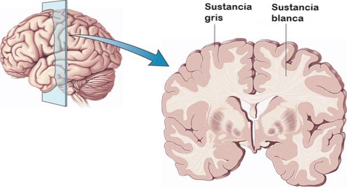
It is usually distinguished from white matter, which is composed of myelinated axons that are responsible for connecting the different areas of gray matter with each other. In general, white matter is the one that gives more speed to information processing.
As myelin has a whitish color, it is seen roughly as a set of white mass (hence its name).
The gray matter occupies approximately 40% of the human brain. The remaining 60% is made up of white matter. However, the gray matter consumes 94% of brain oxygen.
The brain has been advancing phylogenetically in species, reaching its maximum development in humans. The outermost layer or surface of our cerebral cortex is the newest and most complex area. This is covered in a layer of gray matter..
It has been found that the larger the animal, the more complex the substance is and the more convolutions it has. Beneath that layer of gray matter are the myelinated axons of the white matter..
Article index
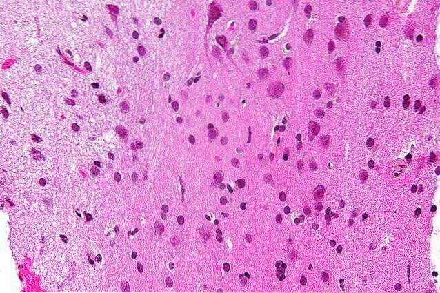
The gray matter mainly includes a dense set of cell bodies, axon terminals, dendrites, etc. Which is known as "neuropil". Specifically, gray matter is made up of:
- Neuron bodies and their bodies. That is, the nuclei of nerve cells.
- Unmyelinated axons. Axons are extensions that extend from neuronal bodies and carry nerve signals.
- Dendrites or small branches arising from an axon.
- Axon terminal buttons, which are the ends of the axons that connect with other nerve cells to exchange information.
- Glial cells or support cells. Specifically two types: astrocytes and oligodendrocytes. This class of cells transport energy and nutrients to neurons, maintaining proper functioning of these and their connections..
- Blood capillaries.
The gray matter may contain some myelinated axons. However, compared to the white matter they are minimal. That is why they are observed in different colors.
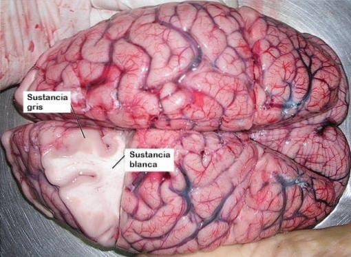
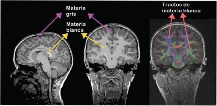
Generally speaking, the gray matter is located mainly on the surface of the brain, while the white matter is in the innermost layers of the cerebral cortex..
In contrast, the opposite pattern is observed in the spinal cord. The gray matter is inside the medulla, surrounded by white matter. In this place, the gray matter takes on the shape of a butterfly or letter "H".
Gray matter has also been found inside the basal ganglia, thalamus, hypothalamus, and cerebellum..
More specifically, we can observe gray matter in:
- The surface of the cerebral hemispheres (cerebral cortex).
- The surface of the cerebellum (cerebellar cortex).
- Deep parts of the cerebellum such as the dentate nucleus, the emboliform, the fastigium and the globose.
- In deep areas of the hypothalamus, thalamus and subthalamus. As well as in the structures that make up the basal ganglia (globus pallidus, putamen and nucleus of accumbens).
- In the brain stem, in structures such as the red nucleus, the nuclei of the olive, the substantia nigra, and the nuclei of the cranial nerves.
- Inside the spinal cord, including the anterior horn, lateral horn, and posterior horn.
Gray matter is found in areas of the brain involved in motor control, sensory perception (sight, hearing), memory, emotions, language, decision-making, and self-control..
The gray matter serves to process and interpret information in the brain and spinal cord. Structures made of gray matter process information from sensory organs or other areas of gray matter.
These signals reach the gray matter through myelinated axons, which make up most of the white matter. Thus, white and gray matter work together.
Additionally, gray matter induces motor signals in your nerve cells to trigger reactions to stimuli..
Ultimately, this substance is related to information processing, but cannot send it quickly. It is the white matter that is linked to the speedy transmission of information.
The gray matter in the spinal cord is divided into several columns. Each of them performs different functions:
- Anterior gray column: It is composed of motor neurons or motor neurons, which are involved in voluntary movements of muscles. They synapse (connect) with interneurons and cell axons that descend the pyramidal pathway. This pathway consists of a group of nerve fibers that participate in voluntary movements.
- Rear gray column: includes the synapses of sensory neurons. These receive sensitive information from the body such as touch, proprioception (perception of our body) and the perception of vibration..
This information comes from receptors located in the skin, bones and joints; and reaches the sensory neurons. These neurons are grouped in the so-called dorsal root ganglia.
Subsequently, these data are sent via axons to the spinal cord through spinal tracts such as the spinothalamic tract and the medial dorsal-lemniscal pathway..
- The side gray column: It is located in the central part of the spinal cord. It only exists in the thoracic and lumbar segments. It has preganglionic neurons of the sympathetic nervous system. The latter is the one that does not prepare for fight or flight reactions by accelerating our heart rate, dilating the pupils and increasing sweating..
Santiago Ramón y Cajal, the Spanish doctor who received the Nobel Prize in Medicine in 1906, studied and classified the neurons of the gray matter.
In the spinal cord, several types of neurons coexist according to the characteristics of their axons:
They are found in the horn or anterior column of the medulla and have different sizes and shapes. Their axons start directly from the nervous system.
Among these are alpha motor neurons and gamma motor neurons..
- Alpha motor neurons: they make direct synapses with muscle fibers. When activated, they can contract muscles. They are large neurons with a stellate soma. Its dendrites are long and have many branching.
- Gamma motor neurons: they connect with the intrafusal muscle fibers. That is, fibers that serve to detect the level of stretching of the muscle and its changes in length. They are smaller than alphas, and also feature a star-shaped soma. They are located between the alpha motor neurons and have numerous dendrites.
- Preganglionic neurons or vegetative protoneuronss: they belong to the autonomic nervous system and are found in the intermediolateral horn. Specifically, at levels D1-L1 and S2-S4. Their nuclei are spindle-shaped, and dendrites depart from their poles. Its axon contains myelin, and it travels to the vegetative ganglia to synapse with other neurons..
They are distributed throughout the gray matter of the medulla. They are multipolar neurons and their soma is star-shaped. The dendrites are short and with several branches. Their axons are part of the white matter, as they are myelinated. These reach the medullary cords of the white matter.
Some of them are sensory neurons. Furthermore, its axons can be ipsilateral (descending from the same side), heterolateral (from the opposite side), commissural, bilateral (from both sides), and pluricordonal (having more than one cord). They can maintain connections with the thalamus and cerebellum.
Also called short axon, they are interneurons scattered throughout the medullary gray matter. They are multipolar neurons, and have a small, stellate soma.
Its axons have multiple branches, connecting with other neurons in the spinal cord. However, they remain within the gray matter.
Although they are not found in the medulla, it has connections to it and that start from it.
The volume of gray matter is a measure of the density of brain cells in a specific part of the central nervous system..
There is a widespread belief that a greater volume of gray matter implies greater intelligence. However, this has been proven to be false. An example is that dolphins have more gray matter volume than humans.
On the contrary, if more than normal density of gray matter is found in the brain, this may mean that the neural connections have not developed correctly. In other words, it could reflect an immature brain..
As the brain develops, many neurons are eliminated by a natural process called "neural pruning." In it, nerve cells and unnecessary connections are destroyed..
This pruning, as well as the maintenance of effective connections, is a symbol of maturity and greater development of cognitive functions..

Yet No Comments