
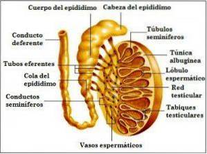
The seminiferous tubules They are tiny channels located in the testicles, where the germination, maturation and transport of the sperm take place towards the testicular network.
The seminiferous tubes occupy between 85 and 90% of the volume of the testes, and fulfill a predominantly exocrine function in the male reproductive system. They are located, specifically, inside the testicular lobes. Each lobe contains between 1 and 5 seminiferous tubes, approximately 70mm long and 0.2mm wide..
Article index
These structures are lined by two types of cells:
These types of cells are found in the walls of the seminiferous tubules, which are composed of several strata.
Basically, these cells produce sperm after going through the processes of mitosis (reproduction of cells) and meiosis (division of cells), respectively..
They are also found inside the seminiferous tubules, enveloping the germ cells.
The sustainable cells of Sertoli complement the nutrition and development of the spermatozoa. They also increase the presence of testosterone in the seminiferous tubules.
For its part, testosterone, which is the male sex hormone, is produced by Leydig cells, which are located in the connective tissue that holds the seminiferous tubules together..
Around the outer surface of the seminiferous tubules is located the tunica propria, also called the limiting layer.
This section is made up of a connective tissue made up, in turn, of myoid cells. These cells, when constrained, make the movement of testicular fluid and sperm through each seminiferous tubule propitious.
Two types of seminiferous tubules are distinguished, depending on the function they fulfill within the testicular structure:
They are coiled in the lobes of the testicular network, and it is within these structures that the spermatogenesis process takes place; that is, the process of sperm formation.
They contribute to the transport of the spermatozoa produced in the convoluted seminiferous tubes, from the mediastinum to the testicular network, also known as rete testis or Haller's network.
This last process is called spermiation. Subsequently, the sperm produced and expelled by the seminiferous tubules are transferred through the testicular network to the vas deferens.
From there, the journey to the epididymis continues, where the spermiogenesis process takes place; that is, the structural formation of the sperm through the allocation of the acrosome.
The acrosome, located in the head of the sperm, in turn contains an important portion of hydrolytic enzymes, essential for the fertilization process.
The seminiferous tubules are extremely important elements within the male reproductive system. If these ducts fail, the formation of sperm, as well as the production of testosterone, would be impossible..
In short, thanks to these small ducts, the sperm production process is feasible, and consequently, the reproductive functions that make fertilization and the generation of life possible among human beings..


Yet No Comments