The Faust technique It is a methodology that allows the concentration of some parasite eggs and / or larvae contained in the feces by flotation. It is used when direct stool tests are negative or when you want to obtain clean samples free of detritus.
Concentration methods for coproparasitological examinations are of three types: by flotation, by sedimentation or by methods that combine the two previous ones. These methods increase the chances of positive results.

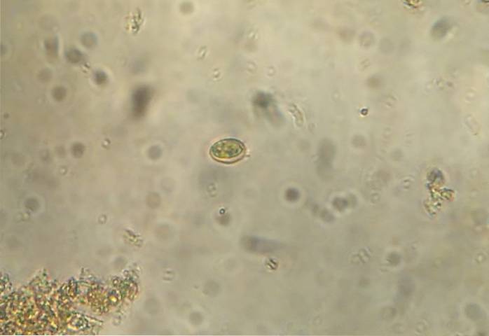
The Faust method consists of mixing part of the stool sample with a substance denser than the eggs or parasites to be concentrated. This causes that, being less dense, they float on the surface. The supernatant fluid is collected and viewed under a microscope for identification and quantification..
This method is used to visualize helminth eggs. In turn, it has proven to be a very sensitive method for the diagnosis of Giardia lamblia, a flagellated protozoan widely spread worldwide. Flotation methods are not recommended for very heavy parasite eggs such as tapeworms and trematodes..
Parasites are one of the most widespread intestinal infections worldwide, especially in poor countries with poor sanitary measures. For this reason, having sensitive methods that allow the identification and quantification of these parasites is very useful for diagnosis and treatment..
Article index
The technique is based on the existence of the different specific gravities of eggs, parasites, cysts, larvae and detritus, using zinc sulfate solutions as a flotation method..
The rationale for the technique is to mix the sample with a zinc sulfate solution that has a higher density than lighter eggs, larvae, or parasites..
This allows the heavier elements to precipitate and the lighter ones to float that appear in the supernatant after centrifugation of the samples..
- Prepare a zinc sulfate solution with a density of 1.18 or 1.2 g / ml if the sample was previously treated.
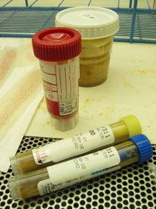
- Prepare a rack with previously labeled test tubes.
- Have a centrifugal machine.
- Have microscope slides and coverslips on hand. All must be labeled
- Ensure availability of a Lugol's solution to stain the sheets.
- Have gauze to filter.
- Have funnels and distilled water.
- Locate labeled plastic or cardboard containers.
- Also applicators and sterile handle of 5 mm.
- A lighter to sterilize the handle.
For any stool examination, the examination begins with what is called the "macroscopic examination" of the samples..
The consistency, color, the presence of what appears to be blood, the presence of mucus, and the presence of adult parasites are described..
Then we proceed to the "microscopic examination" of the stool, this depends on the method. The simplest is the direct smear method, which is the simplest microscopic observation method for parasites..
The procedure involves placing a small amount of the sample directly on a slide. Place several drops of saline solution that should be similar in size to the sample. Mix the saline solution with the stool until a homogeneous mixture is formed. Place a coverslip and examine under the microscope.
The second procedure consists of the Faust flotation method, the original version of which consists of:
1- Place about two grams of feces in a suitable container for this purpose.
2- Add 30 ml of zinc sulfate flotation solution with which an emulsion is made by mixing the solution with the feces.
3- Strain with a metal strainer into a second container and transfer to a test tube.
4- Add more flotation solution until a meniscus forms in the tube.
5- Place a glass coverslip on the meniscus. Let it rest for 10 to 15 minutes.
6- Remove the coverslip and place it on a slide, which will be examined under the microscope.
Originally the method did not use centrifugation, however it is now included as better results are obtained. The technique involves a series of steps to achieve a proper procedure, these are as follows:
1- The feces are washed with water, mixed well and then filtered with gauze folded in four. The sample is placed in a test tube.
2- Centrifuge and remove the supernatant (samples that are kept above water). Steps 1 and 2 are repeated until the supernatant is "clear".
3- Zinc sulfate is added to the filtered and centrifuged sample.
4- Mixes well.
5- Centrifuge again for 1 minute at 2500 rpm (revolutions per minute).
6- The supernatant is recovered with a sterile loop of about 5mm; the tubes should not be shaken.
7- The sample recovered from the supernatant is placed on a slide and a drop of Lugol can be placed to color, the coverslip is placed and observed under a microscope.
8- The containers and test tubes are labeled.
- The elements used for the diagnosis can be seen clean and without “detritus”, this facilitates the observation of the sheet and reduces the time used for the diagnosis.
- In the supernatant, both larvae, eggs and / or cysts are recovered..
- It is a very low cost method.
- The procedure is very simple and easy to implement.
- Diagnosis is fast and accurate.
- Due to the importance and high incidence of parasitosis in poor countries, these low-cost and easy-to-use methods are ideal for the diagnosis and monitoring of these pathologies..
The density of the flotation solution produces a contraction of the larvae, that is, they shrink and, in a very short period, can deform. This forces the examiner to make the diagnosis immediately and the treated samples cannot be kept for future examinations..
As with all microscopic identification methods, it takes a highly experienced examining staff to make accurate diagnoses..
The rapid deformation of the elements necessary for the diagnosis, although they are an obvious disadvantage, can be corrected by making immediate microscopic observations.


Yet No Comments