
The cardiac muscle tissue, Generally called myocardium, it represents the most important tissue component of the heart. Both from the point of view of its size, since it constitutes the majority of the cardiac mass, and its function, since it is the one that develops contractile activity.
The heart also has other types of tissue: a fibrous one that covers it inside (endocardium) and outside (epicardium); another that participates in the separation between the atria and the ventricles; another that separates the atria and ventricles from each other and a valve tissue.
Without ruling out the importance of these fibrous tissues in the cardiac architecture as a support for the mechanical activity of the heart, nor their role in the directionality of the blood (valves), it is the myocardium that generates the electrical and contractile activities of the heart that are essential. for life.
Article index
When talking about tissues, it refers to structures composed of similar cells but that can be of various types and that can be organized in such a way that they work together, resulting in a coordinated function from the physiological point of view.
Cardiac muscle tissue is one of those types of tissue, which, as its name indicates, is muscular in nature, and which fulfills the function of contracting and developing forces that produce displacements of organic components or other external elements..
The characteristics of a tissue can be defined from a structural point of view, both anatomical and histological, and also from a functional point of view. The structure and function of a cell, tissue, organ or system are related.
The structural aspects will be reviewed in the histology section, while here we will refer to some functional characteristics that are grouped under the name of "properties of the heart" and include: chronotropism, inotropism, dromotropism, bathmotropism and lusotropism..
To understand this property, it is necessary to consider that all muscular contraction must be preceded by an electrical excitation in the cell membrane and that it is this excitation that is responsible for triggering the chemical events that will end in mechanical action..
In skeletal muscles, this excitation is the result of the action of a nerve fiber that is in close contact with the muscle cell membrane. When this fiber is excited it releases acetylcholine, an action potential is produced in the membrane and the muscle cell contracts.
In the case of myocardial tissue, the action of a nerve is not required; This tissue has modified cardiac fibers that have the capacity to generate, by themselves, without anything that commands them and automatically, all the excitations that cause cardiac contractions. This is what is called chronotropism.
This property is also called cardiac automatism. The cells that have this capacity for automatism are grouped together in a structure located in the right atrium known as the sinus node. Since this node sets the pace for heart contractions, it is also called a cardiac pacemaker..
Cardiac automatism is the property that allows a heart to continue beating even when removed from the body and what makes heart transplants possible, something that would not have been possible if the reconnection of nerves were required to activate the myocardium..
It refers to the ability of myocardial tissue to generate mechanical force (inos = force). This force is generated because once the cells are excited, molecular phenomena are triggered that shorten the size of the cardiac muscle fibers.
As the ventricular myocardial tissue is organized as surrounding hollow chambers (ventricles) filled with blood, when the muscular walls contract on this blood mass (systole) they increase the pressure in it and move it, directed by the valves, towards the arteries.
Inotropism is like the final objective of cardiac function, since it is this property that constitutes the essence of myocardial tissue, by allowing the movement and circulation of blood to the tissues and from there back to the heart.
It is the ability of the heart muscle to conduct the excitation that originates in the cells of the sinus node, which is the natural pacemaker, and that to be effective on the myocardial cells must reach them in their entirety and practically at the same time.
Some fibers in the atria have specialized in conducting excitation from the sinus node to the contractile myocytes in the ventricle. This system is called the "conduction system" and includes, in addition to ear beams, the bundle of His with its two branches: right and left, and the Purkinje system.
It is the ability of cardiac muscle tissue to respond to electrical stimuli by generating its own electrical excitations, which, in turn, are capable of producing mechanical contractions. Thanks to this property, the installation of artificial pacemakers has been made possible.
It is the ability to relax. At the end of the cardiac contraction, the ventricle is left with a minimum volume of blood and it is necessary for the muscle to relax completely (diastole) so that the ventricle can fill again and have blood for the next systole.
The primary function of the myocardium is related to its ability to generate mechanical forces, which when exerted on the blood mass confined within the ventricles produce increases in its pressure and in its tendency to move towards places where the pressure is lower..
During diastole, when the ventricles are relaxed, pressure in the arteries keeps the valves that communicate with the ventricles closed and the heart fills up. In systole the ventricles contract, the pressure increases and the blood ends up leaving the arteries.
In each contraction, each ventricle pushes a certain amount of blood (70 ml) towards the corresponding artery. This phenomenon is repeated as many times in a minute as the heart rate, that is, the number of times the heart contracts in a minute..
The whole body, even in a state of rest, needs the heart to send it about 5 liters of blood / min. The volume that the heart pumps in one minute is called cardiac output, which is equal to the amount of blood with each contraction (stroke volume) multiplied by the heart rate..
The essential function of the heart muscle is, therefore, to maintain adequate cardiac output so that the body receives the amount of blood necessary for the maintenance of its vital functions. During physical exercise the needs increase and the cardiac output also increases.
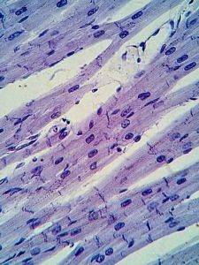
The myocardium has a histological structure very similar to that of skeletal muscle. It is made up of elongated cells about 15 µm in diameter and about 80 µm long. These fibers undergo bifurcations and come into close contact with others, forming chains.
The myocytes or cardiac muscle fibers have a single nucleus and their internal components are organized in such a way that when observed under a light microscope they offer a striated appearance due to the alternating succession of light (I) and dark (A) bands, as in muscle skeletal.
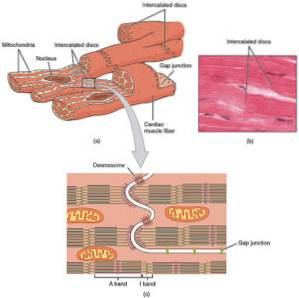
The fibers are made up of a set of thinner and also cylindrical structures called myofibrils, which are arranged along the major (longitudinal) axis of the fibers. Each myofibril results from the sequential union of shorter segments called sarcomeres.
The sarcomere is the anatomical and functional unit of the fiber, it is the space between two Z lines. In them, thin actin filaments are anchored on each side that are directed towards the center of the sarcomere without their ends touching, which interdigitate (intertwine) with thick myosin filaments.
The thick filaments are in the central region of the sarcomere. That area where they are is the one that can be seen, in the light microscope, as the dark band A. From each of the Z lines that delimit a sarcomere to that band A there are only thin filaments and the area is clearer ( I).
Sarcomeres are surrounded by a sarcoplasmic reticulum that stores Ca ++. Invaginations of the cell membrane (T tubes) reach the reticulum. The excitation of the membrane in these tubules opens Ca ++ channels that enter the cell and cause the reticulum to release its Ca ++ and trigger contraction.
Cardiac muscle fibers come into contact with each other at their ends and through structures called intercalary discs. The junction is so tight at these sites that the space separating them is about 20 nm. Here desmosomes and communicating unions are distinguished.
Desmosomes are structures that link one cell to the next and allow the transmission of forces between them. Communicating unions gap junctions) allow ionic flow between two neighboring cells and cause excitation to be transmitted from one cell to another and the tissue to function as a syncytium.


Yet No Comments