The Voges-Proskauer test is a biochemical test used to help identify bacteria belonging to the Enterobacteriaceae family. It is especially useful for differentiating strains of Escherichia coli from Klebsiella and Enterobacter, among other.
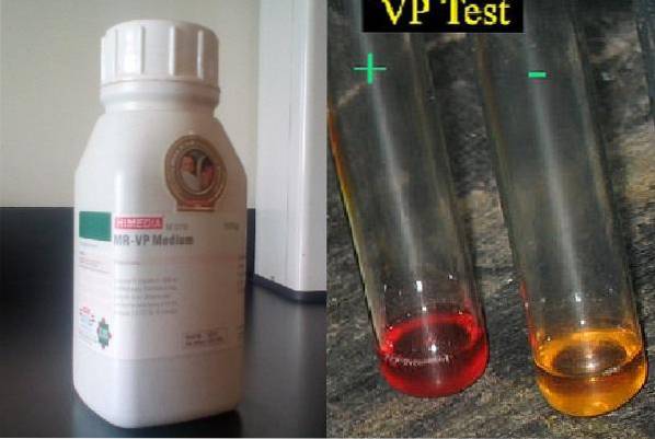
The test is performed in a liquid culture medium called Methyl Red-Voges Proskauer, better known by the acronym RM / VP. This medium is composed of buffered polypeptone, glucose, dipotassium phosphate and distilled water..
The current RM / VP medium is a modification of the Clark and Lubs medium, which originally contained a lower concentration of peptones and glucose. Therefore, less of the hydrogen ion, required for the positive Voges-Proskauer reaction, was produced..
The test is based on the ability of the microorganism to use glucose through the butylene-glycol route, and form a neutral final product called acetoin, in the presence of oxygen and an alkaline pH.
In the RM / VP medium, in addition to being able to reveal the Voges-Proskauer test, the methyl red test can also be revealed.
Article index
The pluripeptones present in the medium provide the essential nutritional requirements for bacterial growth. For its part, glucose is the main compound. Many bacteria have the ability to metabolize glucose and form pyruvic acid.
Pyruvic acid is a midpoint in glucose metabolism and from there each microorganism can take different routes. Some will form mixed acids, such as lactic acid, acetic acid, formic acid, and succinic acid, and others will form neutral products like 2,3-butanediol..
The Voges-Proskauer test reveals the ability of the microorganism to form acetyl methyl carbinol (acetoin), an intermediate product of 2,3-butanediol under aerobic conditions.
Acetoin is reduced and forms 2,3-butanediol, but this reaction is reversible, so if 2,3-butanediol is oxidized, acetoin is formed. Therefore, oxygen is essential.
Dipotassium phosphate is the buffer that buffers the mixture to pH 6.9 ± 0.2.
To demonstrate the reaction, a development must be carried out using two reagents (Barrit reagents), known as Voges A and Voges B.
Voges A is a 5% solution of α-naphthol, and Voges B is a 40% potassium hydroxide preparation. If potassium hydroxide is not available, it can be replaced by 40% sodium hydroxide.
Α-Naphthol is a catalyst that will increase the color intensity of the reaction, making the test more sensitive. The α-naphthol must always be added first, shaking the tube so that the medium comes into contact with the oxygen. In this way, the acetoin present is oxidized to diacetyl, and 2,3-butanediol is oxidized to form acetoin, passing this to diacetyl..
This is how α-naphthol will bind to diacetyl, which in turn has joined the guanidine nucleus present in the amino acid arginine, the latter coming from pluripeptones.
For its part, potassium or sodium hydroxide is responsible for absorbing the COtwo and of reacting with peptones. This reaction causes the formation of a salmon-pink color, clearly visible after shaking the tube very well..
Correct amounts of diacetyl, peptone, and α-naphthol must be mixed for color to occur instantly. If this does not happen, the tube is allowed to rest for 15 minutes before interpreting..
Usually the test is positive after 2 to 5 minutes, when a faint pink color can be seen. If it is left to rest for 30 min to 1 hour the intensity of the color will be maximum (intense red).
A negative test will show up when the broth turns yellow. After 1 hour, if the test is negative, a copper color may form as a result of the reaction of potassium hydroxide on α-naphthol.
Weigh 17 g of the dehydrated culture medium and dissolve in a liter of distilled water. Let stand for 5 minutes. Heat to a boil to dissolve completely. Serve 3 to 4 ml in tubes and sterilize in autoclave at 121 ° C for 15 minutes.
The dehydrated culture medium is beige in color and the prepared medium is light amber in color..
The final pH of the medium is 6.9 ± 0.2.
Weigh out 5 g of α-naphthol and dissolve in 50 ml of ethyl alcohol (absolute). Then continue adding ethyl alcohol until it reaches 100 ml..
Weigh 40 g of potassium hydroxide and dissolve in 50 ml of distilled water in a beaker. The glass must be placed in a cold water bath to control the temperature, because when the preparation is dissolved, the temperature rises sharply.
After the solution is cold, it is transferred to a volumetric flask and made up to 100 mL with distilled water..
To perform the Voges-Proskauer test, an RM / VP broth is inoculated with the microorganism under study, from a pure culture for 18 to 24 hours..
The inoculum should not be very dense. It is incubated at 35-37 ° C for 24 to 48 hours, although incubation for several days is sometimes necessary. Cowan and Steel believe that 5 days is the minimum incubation time necessary to detect all positive Voges-Proskauer (VP) species of the Enterobacteriaceae family..
Separate a 1 mL aliquot into a tube and develop as follows: Place 12 drops (0.6 mL) of Voges A reagent and 4 drops (0.2 mL) of Voges B. Mix to aerate and allow to settle for 5 - 10 minutes before interpreting. However, if the test is still negative, let it sit and observe the tube after 30 minutes to 1 hour..
The appearance of a pinkish-red color indicates that the Voges-Proskauer reaction is positive. If the medium remains yellow the reaction is negative.
Adding developers in the order and quantity indicated is essential to avoid false negatives..
The Voges-Proskauer test is useful to differentiate between strains of E. coli that are VP negative, of the genera Klebsiella, Enterobacter, Serratia, among others, that are VP positive.
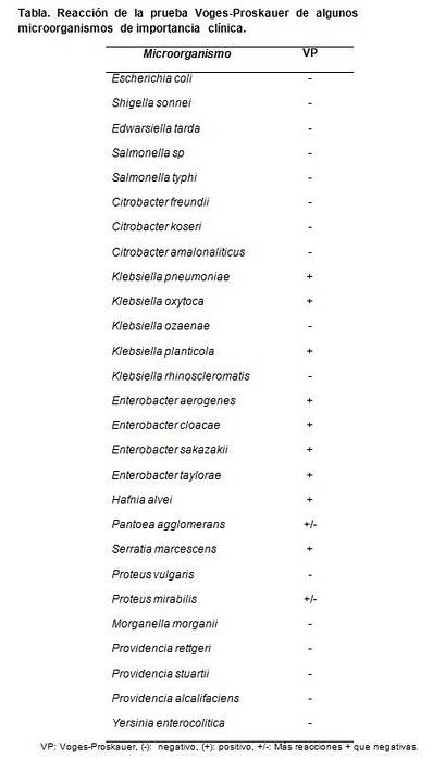
Control strains can be used to test the quality of the prepared medium, including Escherichia coli ATCC 25922, Klebsiella pneumoniae ATCC 700603, Proteus mirabilis ATCC 43071, Salmonella typhimurium and Enterobacter cloacae ATCC 13047.
Expected results are positive Voges-Proskauer reactions only for K. pneumoniae Y E. cloacae. The rest give negative reactions.


Yet No Comments