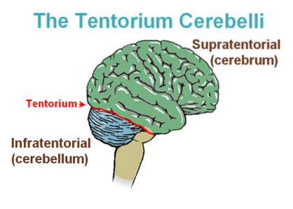
The cerebellum tent It is a thick, semicircular septum shaped like a tent, which covers the cerebellum and separates it from the brain. The lamina that forms the tentorium comes from a prolongation of the dura, the outermost of the meninges, which are the layers that cover the central nervous system (CNS).
It has a fixed edge (which is posterior) and a free edge (which is anterior). The fixed part is convex and inserts into the temporal bone, following a protrusion of the sphenoid bone until it reaches the occiput. For its part, the free edge acquires a concave shape and limits the orifice through which the brainstem opens..
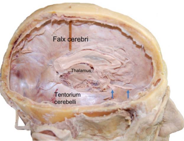
This lamina is located in the posterior cranial fossa and divides the brain space into supratentorial, located over the tentorium, e infratentorial, located below it.
The tent serves as a guide for the doctor when operating on a brain tumor, as different surgical techniques are used if the lesion is above or below the tent.
Article index
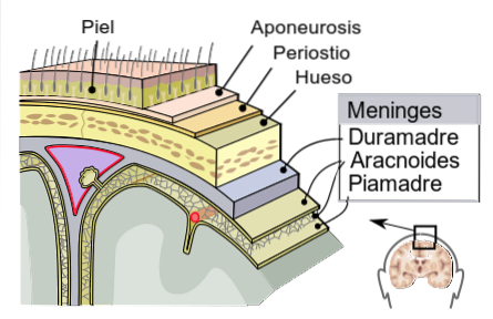
The meninges are three membranes that cover the central nervous system and provide additional protection to that provided by the skeleton. From the inside out they recognize the pia mater, the arachnoid and the dura.
The first two are in close contact and communicated through a rich vascular network. As for the latter, this constitutes the outermost and fibrous layer of the three. It is thick and resistant and forms three specialized partitions from extensions of its own structure.
The sickle of the brain separates the upper part of the two hemispheres of this organ; for its part, the falx cerebellum protects the neurological structure called vermis found between the cerebellar lobes.
The tentorium cerebellum is the second largest reflection of the dura. It is located in the posterior cerebral fossa and separates the cerebellum from the temporal and occipital lobes of the brain.
It was first described in 1732 by the French anatomist Jacques Winslow, who included the term "cerebellum tent" in his publications regarding this structure..
This hard reflection divides the brain space into two parts, supratentorial and infratentorial. The infratentorial is occupied by the cerebellum and the brain troche. Thus, both parts are communicated at the free anterior border of the tentorium, through the tentorial incisure, area through which the brainstem passes.
From the 16th day of gestation, the formation of the primitive central nervous system begins with the migration of cells that will give rise to the brain and spinal cord. Around these structures a cell covering is formed that will give rise to the innermost layer of the meninges..
Around 4ta week, the primitive cerebellum completes its formation and a long cell layer can be seen in the cerebellar spaces that form a middle portion of the fetal cerebellum.
The nuclei of some cranial nerves begin their formation in the 5ta week, and the well-developed primitive dura can now be seen. Covering these elements, a considerable number of cells are observed that will differentiate to form the skull.
Once the fetal cartilaginous skull is formed, by 7ma week of gestation, the primitive dura is completely differentiated and condensed.
The middle portion formed in the 4ta week disappears and the cerebellar tent can be seen in the location it will have after birth.
The tentorium cerebellum runs in an upward direction from back to front and is located at the back of the fossa that houses the brain..
Its anterior border is concave, free of insertions and has a U-shape. It forms the posterior limit of the tentorial incisure, what is the space through which the brain stem passes.
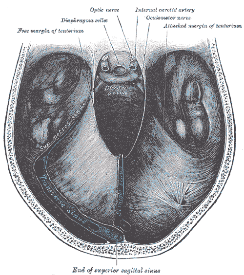
In contrast, the posterior border is convex and fixed. This margin can in turn be divided into two parts, an internal one and a posterior one..
The internal portion is attached to the superior border of the petrous part of the temporal bone, while the posterior portion is attached to the anterior superior aspect of the occipital bone and the parietal bone..
Since its first description in 1732, it is known that the term "tent" is not the most appropriate to describe this fibrous bundle of the dura mater..
Although it is located in the upper part of the cerebellum providing an additional protective layer, this septum fulfills a primary function as a support for the brain.
The cerebellum tent carries about 1,200 grams of brain weight and keeps the brain positioned in the brain fossa..
It also prevents excessive movement of the brain in the case of trauma and deformities of the cerebral lobes.
In addition to this, it separates the brain space into the supra and infratentorial regions, depending on the location above or below the tent, which becomes important in brain surgery..
The technique used in the surgical approach to the brain depends on the location of the structure to be operated on.
To choose the best option, the cerebellum tent is used as an anatomical guide that, in addition to separating the encephalic space, is used as a route of entry to the cerebral elements..
Thus, those lesions located towards the external border of the cerebellum can be approached laterally, while for those located on the medial border, the occipital approach is preferred..
Regarding pathologies, the increase in intracranial pressures caused by space-occupying lesions, such as tumors, hemorrhages or cerebral edema can cause a serious condition known as brain herniation.
Hernia is the protrusion of the brain from one cranial space to another. They are divided into supra or infratentorial.
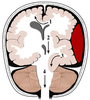
In the supratentorials, one of the most common sites for the exit of the brain is through the tentorial incisura, which is the space limited by the anterior border of the cerebellum, through which the brainstem passes.
On the other hand, in infratentorials the brain exerts great pressure on the tent, causing the cerebellum to protrude through the foramen magnum.
Brain herniation is a clinical and surgical emergency that must be treated immediately, as it can be fatal.


Yet No Comments