
The Kinyoun stain is a staining technique used to stain acid-fast bacteria and parasites. It was born from the modification of the Ziehl-Neelsen coloration; Both techniques are interpreted in the same way but differ in two elements: in the preparation of the main reagent and in that the Kinyoun technique does not use heat.
For this reason it is also known as cold-modified Ziehl-Neelsen or Kinyoun cold stain. It is indicated for the coloring of Mycobacterium tuberculosis, Mycobacterium leprae, atypical mycobacteria, Nocardias sp, Cryptosporidium parvum, Cryptosporidium meleagridis, Cryptosporidium felis, Cryptosporidium muris Y Cyclosporas cayetanensis.
It is worth noting that Nocardias stain weakly with this technique since they are partially acid-alcohol resistant, so for this genus there is a modification of the methodology.
In turn, the cold Kinyoun technique has been combined with the trichrome technique modified by Didier for the detection of coccidia (Cryptosporidium parvum and Isospora belli) and microsporidia spores (Enterocytozoon bieneusi Y Encephalitozoon intestinalis).
Article index
The main staining reagent is carbolfuchsin or carbol fuchsin, which has the property of binding to the carbolic acids existing within the waxy cell wall, rich in lipids (mycolic acids) of mycobacteria and certain parasites..
This bond is not counteracted by the acid bleach; therefore, microorganisms are defined as acid-alcohol resistant.
Unlike the Ziehl-Neelsen technique -which fixes the dye through heat-, in the Kinyoun technique this step is not necessary, since the carbol fuchsin solution prepared for this technique contains a high concentration of phenol..
Phenol dissolves the lipid material in the cell wall, allowing the carbolfuchsin dye to enter. After the dye penetrates, it remains fixed despite washing with the acid alcohol.
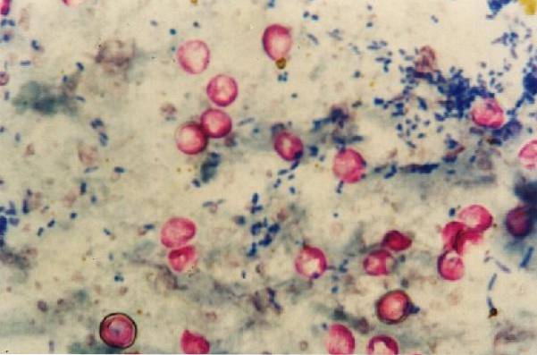
In this way, acid-resistant microorganisms take on the characteristic red color, while everything that is not acid-resistant alcohol becomes discolored and stained blue..
- Modified carbol fuchsin.
- Alcohol-acid.
- Methylene blue.
- Basic fuchsin: 4 gr.
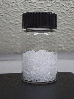
- Phenol: 8 ml.
- Alcohol (95%): 20 ml.
- Distilled water: 100 ml.
The basic fuchsin should be dissolved slowly in the alcohol, mixing constantly. Subsequently, the crystallized phenol is melted in a water bath at 56 ° C. Once dissolved, 8 ml are added to the fuchsin solution prepared above.
- Concentrated hydrochloric acid: 3 ml.
- Ethanol (95%): 97 ml.
It must be measured, joined and mixed.
- Methylene blue: 0.3 g.
- Distilled water: 100 ml.
It is weighed and dissolved.
1- Prepare a smear directly from the sample, which can be sputum, lung fluid, urine sediment, cerebrospinal fluid or feces, among others; or from a suspension of microorganisms obtained from pure colonies developed in primary culture media.
2- Fix the smear with heat.
3- Place the smear on the staining bridge and cover with the prepared Kinyoun carbol fuchsin reagent. Let it rest for 3 or 5 minutes.
4- Wash with distilled water.
5- Bleach with acidic alcohol for 3 minutes and wash again with distilled water.
6- Bleach again with acidic alcohol for 1 or 2 minutes until no more coloring is carried away.
7- Wash with distilled water and allow to drain, placing the slide in a vertical position.
8- Cover the preparation with methylene blue and leave to act for 4 minutes.
9- Wash with distilled water and allow to air dry.
10- Examine at 40X and then at 100X.
If you want to improve and speed up the staining of acid-fast microorganisms, add 1 drop of a wetting agent (such as Tergitol No. 7) to 30 or 40 ml of Kinyoun Carbol Fuchsin.
Some labs change the methylene blue contrast dye to bright green or picric acid; the first gives a green color to the background and the second generates a yellow color.
To improve the staining of bacteria of the genus Nocardia, a modification of the Kinyoun stain is used. The technique is as follows:
1- Cover the smear with Kinyoun carbol fuchsin for 3 minutes.
2- Wash with distilled water.
3- Briefly discolor with acidic alcohol prepared at 3% until no more coloring is carried away.
4- Wash again with distilled water.
5- Cover the preparation with methylene blue and leave for 30 seconds.
6- Wash with distilled water and allow to air dry.
This technique is recommended for the analysis of stool samples for coccidia and spores of Microsporidium sp at the same time. The procedure to follow is as follows:
1- Cover the smear with Kinyoun carbol fuchsin for 10 minutes.
2- Remove the dye and wash with distilled water.
3- Bleach for 30 seconds with hydrochloric acid alcohol.
4- Wash again with distilled water.
5- Cover the smear with trichrome solution for 30 minutes at 37 ° C.
6- Wash with distilled water.
7- Bleach for 10 seconds with acetic acid alcohol.
8- Wash the smear for 30 seconds using 95% ethanol.
As a positive control, smears are prepared with strains of Mycobacterium tuberculosis and stain with the prepared reagents to verify that the bacteria take the proper color (red-fuchsia).
Negative controls can also be used preparing smears with any strain that is not acid-fast, thus verifying that the entire sample takes on the contrasting color..
The Kinyoun technique is simpler since it eliminates the heating step, but its main advantage is that it avoids the emission of vapors, highly toxic and cancer-causing in the long term. Therefore, the Kinyoun stain is safer for staining personnel..
It is important to take into account that care must be taken that the reagents do not come into direct contact with the skin, as they are corrosive and the bleach is flammable..
Regarding the disadvantages, a negative smear does not necessarily indicate that the organism is not present. In addition, the presence of cellular debris can lead to false positives, leading to confusion in the diagnosis..


Yet No Comments