
The types of neurons The main factors can be classified according to the transmission of the impulse, the function, the direction, by the action in other neurons, by their discharge pattern, by the production of neurotransmitters, by the polarity, according to the distance between axon and soma, according to the morphology dendrites and depending on the location and shape.
There are approximately 100 billion neurons in our brain. On the other hand, if we talk about glial cells (those that serve as support for neurons), the number increases to about 360 billion.
Neurons resemble other cells, among other things, in that they have a membrane that surrounds them, contain genes, cytoplasm, mitochondria, and trigger essential cellular processes such as synthesizing proteins and producing energy.
But, unlike other cells, neurons have dendrites and axons that communicate with each other by electrochemical processes, establish synapses, and contain neurotransmitters..
These cells are organized as if they were trees in a dense forest, where their branches and roots intertwine. Like trees, each individual neuron has a common structure, but its shape and size vary..
The smallest can have a cell body only 4 microns wide, while the cell bodies of the largest neurons can be as wide as 100 microns. In fact, scientists are still investigating brain cells and discovering new structures, functions, and ways to classify them..
Article index
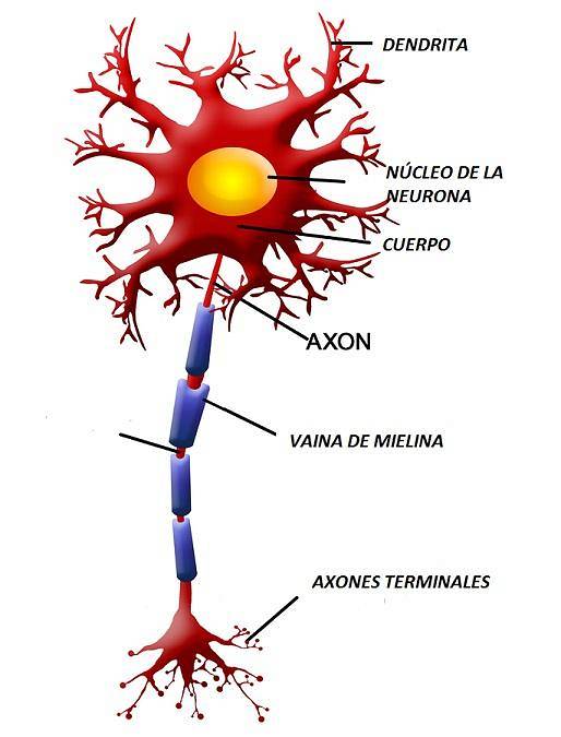
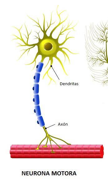
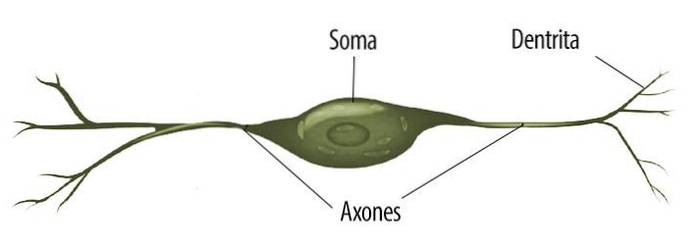
The basic shape of a neuron is made up of 3 parts:
- The cell body: contains the nucleus of the neuron, which is where genetic information is stored.
- The axon: is an extension that works as a cable, and is responsible for transmitting electrical signals (action potentials) from the cell body to other neurons.
- Dendrites: they are small branches that capture the electrical signals emitted by other neurons.
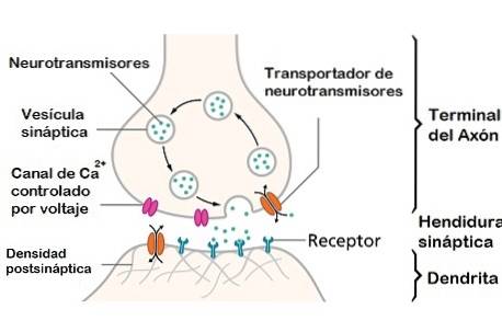
Each neuron can make connections to up to 1000 other neurons. However, as the researcher Santiago Ramón y Cajal stated, the neuronal ends do not merge, but there are small spaces (called synaptic clefts). This exchange of information between neurons is called synapses (Jabr, 2012).
Here we explain the functions and characteristics of up to 35 types of neurons. To make them easier to understand, we have classified them according to different ways.
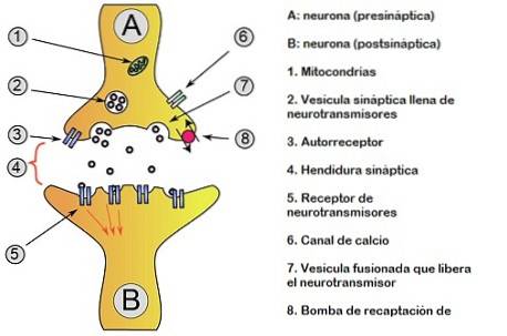
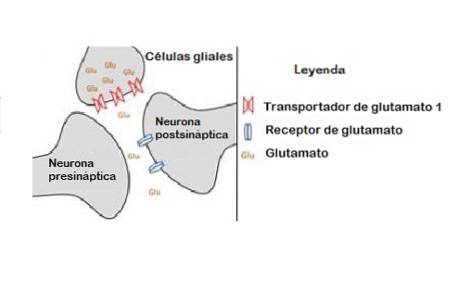
A main classification that we are going to find very frequently to understand certain neuronal processes is to distinguish between the presynaptic neuron and the postsynaptic neuron:
It must be clarified that this differentiation is applied within a specific context and moment.
Neurons can be classified according to the tasks they perform. According to Jabr (2012), in a very common way we will find a division between:
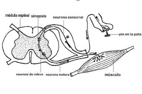
They are those that handle information from the sensory organs: the skin, eyes, ears, nose, etc..
Its task is to send signals from the brain and spinal cord to the muscles. They are primarily responsible for controlling movement.
They act as a bridge between two neurons. They can have longer or shorter axons, depending on how far these neurons are from each other.
They release hormones and other substances, some of these neurons are found in the hypothalamus.
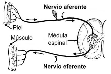
It is another way of calling motor neurons, pointing out that the direction of information transmission is opposite to afferents (they send data from the nervous system to the effector cells).
One neuron influences the others by releasing different types of neurotransmitters that bind to specialized chemical receptors. To make this more understandable, we can say that a neurotransmitter works as if it were a key and the receptor would be like a door that blocks the passage.
Applied to our case it is somewhat more complex, since the same type of "key" can open many different types of "locks". This classification is based on the effect they cause on other neurons:
They are the ones that release glutamate. They are so called because, when this substance is captured by the receptors, there is an increase in the firing rate of the neuron that receives it..
They release GABA, a type of neurotransmitter that has inhibitory effects. This is because it reduces the firing rate of the neuron that captures it..
They do not have a direct effect, but in the long term change small structural aspects of nerve cells.
Approximately 90% of neurons release glutamate or GABA, so this classification includes the vast majority of neurons. The rest have specific functions according to the objectives they present.
For example, some neurons secrete glycine, exerting an inhibitory effect. In turn, there are motor neurons in the spinal cord that release acetylcholine and provide an excitatory result..
Anyway, it should be noted that this is not so simple. That is, a single neuron that releases one type of neurotransmitter can have both excitatory and inhibitory effects, and even modulators on other neurons. Rather, this seems to depend on the type of receptors activated on postsynaptic neurons..

We can pigeonhole neurons by electrophysiological traits.
Refers to neurons that are constantly active.
They are the ones that are activated in bursts.
These neurons stand out for their high firing rates, that is, they fire very frequently. Cells of the globe pallidus, ganglion cells of the retina, or some classes of cortical inhibitory interneurons would be good examples..
This type of neuron releases acetylcholine in the synaptic cleft.
They release GABA.
They release dopamine, which is linked to mood and behavior.
They are those that release serotonin, which can act both by exciting and inhibiting. Its lack has traditionally been linked to depression.
Neurons can be classified according to the number of processes that join the cell body or soma, and can be:
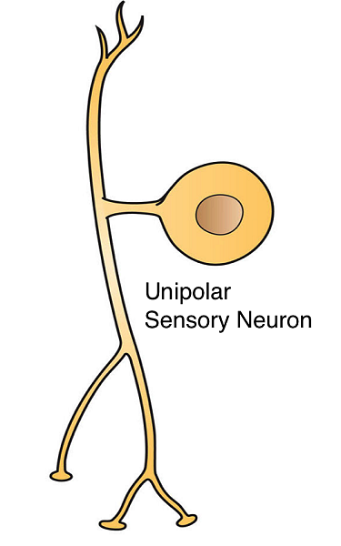
They are those that have a single protoplasmic process (only a primary extension or projection). Structurally, it is observed that the cell body is located on one side of the axon, transmitting the impulses without the signals passing through the soma. They are typical of invertebrates, although we can also find them in the retina.
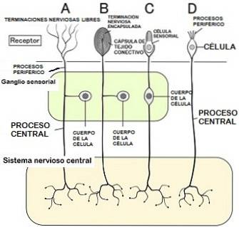
They are distinguished from unipolar ones in that the axon is divided into two branches, generally one goes towards a peripheral structure and the other goes towards the central nervous system. They are important in the sense of touch. Actually, they could be considered a variant of the bipolar ones.
In contrast to the previous type, these neurons have two extensions that start from the cell soma. They are common in the sensory pathways of sight, hearing, smell and taste, as well as vestibular function..

Most neurons belong to this type, which is characterized by having a single axon, usually long, and many dendrites. These can originate directly from the soma, assuming an important exchange of information with other neurons. They can be subdivided into two classes:
a) Golgi I: long axons, typical of pyramidal cells and Purkinje cells.
b) Golgi II: short axons, typical of granule cells.
In this type, dendrites cannot be differentiated from axons, and they are also very small..
In these neurons the axon may be more or less branched, however, it is not excessively far from the body of the neuron (soma).
Despite the number of branches, the axon extends a long distance and remarkably moves away from the neuronal soma.
Its dendrites depend on the type of neuron it is (if we classify it according to its location in the nervous system and its characteristic shape, see below). Good examples are Purkinje cells and pyramidal cells..
This class of neuron has dendrites that divide in such a way that the daughter branches exceed the mother branches in length..
They have traits that are not typical of dendrites, such as having very few spines or dendrites without branching.
There are a multitude of neurons in our brain that have a unique structure and it is not an easy task to classify them with this criterion.
Depending on the shape, they can be considered:
If we take into account both the location and the shape of neurons, we can further refine and detail this distinction:
They are so called because the somas have a triangular pyramid shape and are found in the prefrontal cortex..
They are large pyramidal-shaped motor neurons that are located in the fifth layer of the gray matter in the primary motor cortex..
They are cortical interneurons that are located in the cortex and in the cerebellum.
Tree-shaped neurons found in the cerebellum.
They make up the majority of neurons in the human brain. They are characterized by having very small cell bodies (they are of the Golgi II type) and are located in the granular layer of the cerebellum, the dentate gyrus of the hippocampus and the olfactory bulb, among others..
Named for their discoverer, they are inhibitory sensory interneurons located in the cerebellum (just below the Purkinje cell layer)..
They are considered a special type of GABAergic cell that represents approximately 95% of the neurons of the striatum in humans..
These neurons are inhibitory interneurons of the spinal cord that are connected at their ends with alpha motor neurons, neurons with both ends linked to alpha motor neurons.
They consist of a type of glutamatergic interneurons that are located in the granular layer of the cerebellar cortex and in the cochlear nucleus. Its name is due to the fact that it has a single dendrite that ends in a brush shape.
They are named for the motor neurons located in the spinal cord.
Also called Von Economo neurons, they are characterized by being fusiform, that is, their shape looks like an elongated tube that becomes narrow at the ends. They are located in very restricted areas: the insula, the anterior cingulate gyrus and, in humans, in the dorsolateral prefrontal cortex.
We can affirm that almost all the neurons of the nervous system can be pigeonholed into the categories that we offer here, especially the broader ones. However, it is necessary to point out the immense complexity of our nervous system and all the advances that remain to be discovered in this area..
There is still research focused on distinguishing the most subtle differences between neurons, in order to learn more about the functioning of the brain and associated diseases.
Neurons are distinguished from each other by structural, genetic and functional aspects, as well as the way they interact with other cells. It is even important to know that there is no agreement among scientists when determining an exact number of types of neurons, but it could be more than 200 types.
A very useful resource to learn more about the cell types of the nervous system is Neuro Morpho, a database in which the different neurons are digitally reconstructed and can be explored according to species, cell types, brain regions, etc. (Jabr, 2012)
In summary, the classification of neurons into different classes has been considerably debated since the beginning of modern neuroscience. However, this question can be gradually unraveled, as experimental advances are accelerating the pace of data collection on neural mechanisms. Thus, every day we are one step closer to knowing the totality of brain function.


Yet No Comments