The thoracentesis is a surgical technique in which the chest is punctured to evacuate fluid or drain trapped air. From greek thorako ("Chest") and kehesis (“Perforation”), is understood to be the controlled perforation of the chest for therapeutic or diagnostic purposes.
It is also known as thoracentesis, thoracic paracentesis, or pleurocentesis. This last term is the most correct, since the true purpose of the procedure is to cross the pleura at some specific anatomical point to allow air or fluid to escape that should not be in the pleural space..
It was first carried out in 1850 by Morrill Wyman, an American physician and sociologist, although its formal description was carried out by Henry Ingersoll Bowditch, a prominent Massachusetts physician and abolitionist, remembered not only for his medical achievements but for his radical support of runaway slaves.
Article index
Thoracentesis has two fundamental indications: diagnostic and therapeutic..
When unexplained fluid is evident in the pleural cavity, thoracentesis may be indicated..
By performing the procedure correctly, you will get enough fluid to perform a series of tests. Most cases of pleural effusion are due to infections, cancer, heart failure, and recent thoracic surgeries.
When the presence of fluid in the pleural cavity causes significant discomfort in the patient, thoracentesis can relieve symptoms.
Although it is not the ideal technique to drain massive amounts of fluid, about 1 or 2 liters can be removed, which greatly improves the person's breathing capacity and comfort.
This procedure can be performed by a well-trained physician or an experienced interventional radiologist. In the latter case, they are usually supported by imaging equipment such as ultrasound scanners or tomographs, which significantly reduces the risks of complications..
Whether it is a real-time image-guided thoracentesis or not, the procedure is very similar. There is a technique for draining fluids and another technique for draining air..
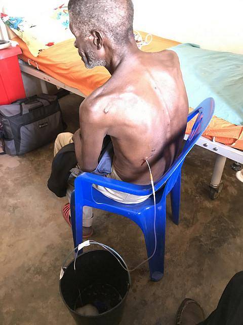
The ideal position of the patient to perform the procedure is sitting. You should drop your shoulders and rest your arms on a table.
The head lowered rests on the arms or with the chin against the chest. The person should be advised to hold their breath to avoid puncturing the lung.
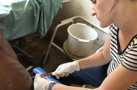
The ideal location of the needle is in the mid-axillary line, between the sixth and eighth intercostal spaces of the affected hemithorax. The approach is made to the back of the patient after asepsis and antisepsis. It is always advisable to infiltrate local anesthetic in the area to be punctured. All materials used must have guaranteed sterility.
The puncture is performed by leaning on the upper edge of the lower rib that forms the selected intercostal space. It is done this way to avoid the vessels and nerves that run along the lower edge of the costal arches. When obtaining fluid, the needle should be connected to a drainage system or manual removal with a large syringe.
Thoracentesis also works to drain air trapped in the pleural space. This phenomenon is known as a tension pneumothorax and can cause dyspnea, hypotension, and cyanosis. The purpose of the technique is to extract the air present between the pleura and the costal wall, preventing it from re-entering.
This procedure is performed with a 10 cc or larger syringe, a three-way stopcock, a guiding catheter, and a one-way flow air valve or Heimlich valve, which can be replaced by a glove finger sealed around the the needle as a craft.
Under standards of asepsis and antisepsis, and with infiltrative local anesthesia, the second intercostal space is punctured on the midclavicular line with the needle connected to the syringe and the valve. Sudden release of air through the system should be felt and immediate patient relief.
Potential complications after thoracentesis are:
Thoracentesis is always painful. It is the job of the person who performs the procedure to try to make it as painless as possible through the use of local anesthetics and a refined technique..
The most intense pain is felt by the patient when the subcostal neurovascular bundle is manipulated. Therefore, thoracentesis should be done with caution..
When the lung is punctured during the procedure, a pneumothorax is likely to occur. It is usually marginal, but sometimes it is more extensive and even massive.
To avoid this, as mentioned above, the patient should be asked to hold their breath at the time of the puncture. May require thoracotomy and permanent drainage.
Although rare, it is one of the most feared complications of thoracentesis due to its difficult management and potential fatality. It occurs when the lung is punctured along with a blood vessel.
The most affected vessels are the subcostals due to poor technique or poor patient cooperation. May need corrective surgery and chest tube placement.
The presence of blood in the pleural space without being accompanied by air is due to subcutaneous or subcostal vascular damage, with compensation of the lung.
Cases of massive hemothorax after subcostal artery damage have been described. The best prevention is an impeccable technique and, if necessary, sedate the patient.
Dyspnea is common during or after thoracentesis. It is related to the re-expansion of the lung and certain local nervous stimuli. If the respiratory distress is very severe, the presence of pneumothorax, hemothorax or hemopneumothorax should be suspected..
Sudden expansion of the affected lung can cause pulmonary edema. The inflammatory response may be the cause of this complication, as it is a damaged lung. Usually self-limiting, although intravenous steroids and oxygen support may be needed for a time.
Stimulation of the vagus nerve that occurs after expansion of the affected lung can cause hypotension and syncope.
It can also be accompanied by nausea, vomiting, paleness, and dizziness. This effect is temporary, but to avoid it it is recommended not to drain more than 1 liter per procedure and to do it slowly.
Local hematomas, seromas, pleural infections, subcutaneous emphysema, cough, inadvertent puncture of the liver or spleen, and anxiety may occur..

Yet No Comments