Toxascaris leonina it is a worm belonging to the phylum Nematoda. It is characterized by its cylindrical shape and its head shaped like an arrowhead. It is an endoparasite, that is, it lives inside its hosts.
Mainly, the living beings in which it likes to stay are cats and dogs, although they can also be hosts of foxes and some other mammals, although in a very small proportion.

This parasite, together with Toxocara cati Y Toxocara canis they are responsible for an infection known as toxocariasis, which affects its hosts. Humans can occasionally become infected by ingesting parasite eggs, either by ingesting contaminated food or water, or by contact with pet feces..
Toxocariasis is an easy disease to treat, but if it is not treated in time, it can lead to the degeneration and gradual and chronic deterioration of different organs of the body..
Article index
The taxonomic classification of Toxascaris leonina is the next:
- Domain: Eukarya
- Animalia Kingdom
- Phylum: Nematoda
- Class: Secernentea
- Order: Ascaridia
- Family: Toxocaridae
- Gender: Toxascaris
- Species: Toxascaris leonina
Toxascaris leonina it is an organism considered eukaryotic, multicellular, triblastic and pseudocoelomates.
The cells of this parasite have a cellular organelle known as the nucleus, within which the DNA is found, well packaged, conforming to the chromosomes. Likewise, these cells are specialized in different functions, such as the absorption of nutrients, the production of gametes and the transmission of nerve impulses, among others..
During its embryonic development process, the three germ layers are present: ectoderm, endoderm and mesoderm. The cells of each layer differentiate into different types of cells, thus forming each of the tissues and organs that will make up the adult worm..
In addition to this, they present an internal cavity known as a pseudocoelom, whose origin is not mesodermal..
These animals present bilateral symmetry, which means that if an imaginary line is drawn along the longitudinal axis of the animal, two exactly equal halves will be obtained..
Their lifestyle is parasitic, which means that they must be inside a host in order to survive, the most common being dogs and cats, although it can also develop in other mammals such as foxes and coyotes, among others..
Toxascaris leonina It is a nematode worm and, as such, it has an elongated, cylindrical shape. They present sexual dimorphism, for which there are well marked morphological differences between female and male specimens.
Females are much taller than males. They can reach up to 10 cm in length and 2 mm in thickness. While the males measure only up to about 6 cm approximately.
The head end of the worm has a kind of cervical fins, which give an arrowhead appearance to the animal's head. At this same end, is the orifice of the mouth, which is surrounded by three lips.
The terminal caudal part of the male has extensions called spicules, which are approximately 1.5 mm long. They are used for the copulation process.
The life cycle of Toxascaris leonina it is quite simple, much less complex than that of other nematodes. Generally, it does not require intermediate hosts or vectors, but when it enters the body of its definitive host, its development ends there..
Sometimes some animals such as certain rodents can intervene in the life cycle as an intermediate host.
The eggs are released to the external environment through the feces. There the larvae undergo certain transformations from a harmless state to an infectious form.
This process depends entirely on the environmental conditions that exist. For example, the ideal temperature for the larvae to molt is 37 ° C, above this the larvae lose their ability to transform. While, at lower temperatures, they can transform but at much slower speeds.
The time required for the larvae inside the eggs to transform and become infective is approximately between 3 and 6 days.
The definitive host, which is generally a cat, dog or also a fox, becomes infected by ingesting food or water that is contaminated with eggs. These pass directly to the stomach of the animal and later to the small intestine.
Once there, the eggs hatch, releasing the infective larvae that were inside them. In the intestine, the larva penetrates the mucosa and intestinal wall and undergoes other transformations inside it until it becomes an adult individual..
Once converted into adult worms, the parasites migrate again towards the intestinal lumen and there the reproduction process takes place, through which the female lays the eggs. These are released to the outside through the feces, to start a new cycle.
This is the regular life cycle of Toxascaris leonina. However, there are times when the eggs are ingested by an intermediate host, such as a rat..
In this case, the eggs hatch in the intestine of the animal, but the larvae do not stay there, but instead initiate a process of migration through the different tissues of the animal and there they remain waiting for it to be ingested by one of its definitive guests.
When the rodent is ingested by a cat, for example, the larvae pass from the animal's tissues into its digestive tract, thus continuing its development, transforming into adult worms ready to lay eggs and continue the cycle..
It is important to note that unsanitary conditions are those that allow the biological cycle of this parasite to run its course, especially when the infected are domestic animals..
With these it is necessary to follow the same hygiene and food safety measures that are followed with the food and water of the rest of the family. This in order to avoid the transmission of certain pathologies.
Toxascaris leonina It is a pathogenic parasite that can cause an infection in its host known as toxocariasis. This mainly affects the host animals of the parasite. However, human beings, especially children, are also susceptible to becoming infected and developing certain symptoms..
In the case of domestic animals, the symptoms that may occur are the following:
- Loss of appetite
- Apathy
- Bristly or disheveled hair
- Weight loss, caused by decreased food intake
- Vomiting that may sometimes contain adult worms
- Globose belly, generated by the accumulation of parasites in the intestine
When humans are infected, either by consuming raw meat or by being in contact with sand infected with animal feces, the following symptoms become evident:
- High fever that can exceed 39.5 ° C
- Swelling of the different groups of lymph nodes in the body
- Loss of appetite
- Generalized chronic fatigue
- Chronic severe pain in the joints
However, in humans, larvae generally do not remain in the intestine, but instead migrate to different organs and cause damage to them, which in turn generate certain symptoms such as:
- Hepatomegaly (enlargement of the liver)
- Inflammation of the liver
- Pneumonitis
- Difficulty breathing
- Chronic cough
- Pneumonia
- Skin problems: rashes, chronic itching, eczema,
- Inflammation of the myocardium
- Endocarditis
- Inflammation of the kidneys
- Alteration of blood values: increase in eosinophils, dysfunction in liver hormones.
These symptoms depend on the organ to which the larvae migrate.
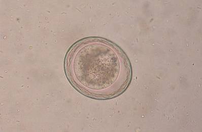
This disease can be diagnosed through three mechanisms: direct observation of stools, blood tests, and imaging tests..
The initial diagnosis of infection by Toxascaris leonina It is diagnosed primarily by looking at stool under a microscope. By observing them it is possible to determine whether or not there is presence of parasite eggs. Likewise, if the parasitosis is very accentuated, adult worms can also be observed in the animal's feces..
Likewise, through blood tests, an infection by Toxascaris leonina. Through these tests, the antibodies that the body synthesizes against these parasites can be identified.
A serological test called ELISA seeks to detect excretion and secretion antigens of second stage larvae (L2), as well as Immunoglobulin G (IgG).
When it is suspected that a person may suffer from a parasitic infection, a magnetic resonance imaging (MRI) or a computed tomography (CT) scan can be performed in which lesions in some organs that are known to be caused by the parasite can be identified.
Because the infection is caused by a nematode parasite, the indicated treatment, in general, is the administration of drugs known as anthelmintics..
The anthelmintics that have been shown to be most effective in treating these types of infections are albendazole and mebendazole. The mechanism of action of these drugs is based on the fact that it causes degeneration in the animal's tissues, mainly at the level of its integument and intestine..
Subsequently, a progressive degeneration occurs in its cytoplasmic organelles. These prevent certain processes such as cellular respiration, which is the one that generates the greatest amount of energy (in the form of ATP molecules)..
By not having the necessary energy production, the parasite ends up remaining totally immobile, until it finally dies. This occurs both in the adult form of the parasite and in its larval stages..
For the rest of the clinical manifestations of the disease, the specialist doctor prescribes the treatment he deems necessary, according to the severity of the symptoms and signs..


Yet No Comments