
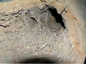
The trabeculae are small sheets found in a type of bone tissue called cancellous bone or trabecular or areolar bone. The trabeculae are arranged irregularly, creating partitions and spaces, adopting the shape of the surface of a sponge.
Although the term is often used to refer to bone tissue, its use is not exclusive to refer to bones. The importance of the trabecular network in bone is that in the spaces that form between the trabecular septa is the bone marrow.
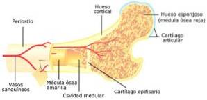
The bone marrow is a specialized structure found in long bones. From the spongy bone tissue begins the formation of erythrocytes, leukocytes and platelets, which are the main cells that make up the blood.
The process of making blood cells from bone marrow tissue is called hematopoiesis.
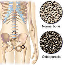
The spongy bone of the pelvis is the site where bone marrow samples are taken to diagnose malignant diseases such as leukemia. Osteoporosis is one of the main pathologies that affect this type of bone tissue, causing a significant deterioration in the surface of the trabeculae.
Article index
The spaces formed by the trabeculae in the spongy bone contain the bone marrow, which is the structure of the body responsible for producing undifferentiated blood cells, allowing them to differentiate and depositing them in the circulation. This process is called hematopoiesis.
In the adult, hematopoiesis only occurs in the bone marrow. On the contrary, during the fetal stage the location of stem cells varies and, therefore, the site at which hematopoiesis takes place also varies..
During the first trimester it occurs in the yolk sac; in the liver and spleen by the second trimester and finally in the bone marrow towards the end of gestation.
The wall that contains the bone marrow is made up of thin, smooth trabeculae with wide spaces. These spaces communicate by contiguity or through delicate channels formed by the junction between trabeculae.
Hematopoiesis begins with the multipotential cell called stem cell. The term multipotential is used since they are cells that have the ability to differentiate into any of the blood cell types.
Erythrocytes, leukocytes, and platelets are the main blood cells that are formed from stem cells. Each cell line develops depending on the trabecular space in which it is found.
So, the location of the stem cells within the cancellous bone determines the type of cell into which it will differentiate..
Blood vessels penetrate into the trabecular spaces, allowing cell and nutrient exchange from bone with the bloodstream.
Bones are made up of a special type of tissue made up of calcium known as woven bone.
The set of bones united by cartilage and ligaments forms the human skeleton, which performs functions of movement, maintenance of posture, containment and protection of the organs..

In addition, bones are the body's main storage site for calcium and phosphate; have an important reserve of fat cells and some contain spaces in which are immature blood cells that continually develop and incorporate new essential components of the blood into the circulation.
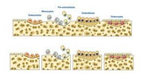
Each bone has a complex structure made up of cells that are restored from time to time, through the formation and elimination of bone tissue. These cells are called osteoblasts Y osteoclasts respectively.
The process through which mature bone cells make and reabsorb bone tissue is known as osseous remodeling.
Bone is the only tissue in the body that has the ability to regenerate with a structure exactly the same as the original and not with scar tissue. When an individual suffers a fracture, bone cells are responsible for forming new tissue that ends up joining the ends of the fractured part.
The balance of osteoblast and osteoclast function is essential for the proper maintenance of this tissue. If any of these cells fail to do their job, there is an increased metabolism in the bone that can lead to wear and tear or abnormal growth..
For example, when there is increased bone resorption by osteoclasts, without the corresponding formation of new cells, there will be loss of bone tissue. This pathology is known as osteoporosis.
In general, there are two types of bone tissue that is found in all bones but distributed differently in each one. These are the compact tissue and the spongy tissue.
Although both share essential characteristics, their structures and functions as well as their response to trauma, are completely different.
Compact bone is a hard and dense structure, very resistant to compression, which is located mainly in the body of the bones. It is organized in several layers of concentric tissue that surrounds a main channel that supplies it with blood. This area is called Havers canal.
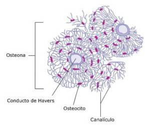
This type of tissue contains a specialized vascular system as well as hormone receptors that regulate the storage and distribution of calcium and phosphate..
The set that forms the main Haversian canal with the complicated network of canals, ducts and spaces through which the bone is nourished, is called osteone or Havers system. Osteon is considered the structural unit of compact bone.
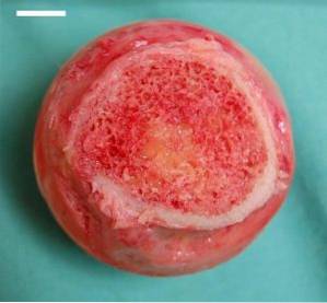
Cancellous bone does not contain osteons but rather has hollow spaces that form an elastic and resistant structure that cushions weight well. Its name comes from the shape it takes, similar to a sponge.
It is located mainly in the upper and lower extremities of the long bones and inside the rest of the bones.
Within this type of tissue there are lamellae arranged in an organized manner called trabeculae..
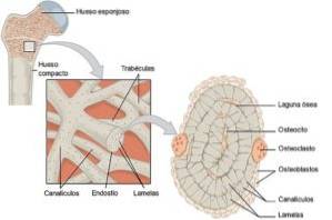
The trabeculae form small partitions that create spaces inside the bone. Depending on the arrangement of these septa, up to three different types of cancellous bone can be distinguished.
Inside the trabecular spaces is the bone marrow, which is a tissue that is part of the blood system and is responsible for forming the precursor elements of blood cells.
Cancellous bone tissue has a greater surface area for cell turnover and regeneration than compact tissue. In addition, it contains the bone marrow. For these reasons, bone pathologies can be observed frequently in this part of the tissue..
The osteoporosis is a common disease especially in postmenopausal women and the elderly, in which there is an imbalance between the formation and resorption of the bone in which resorption predominates.
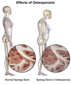
The first radiological signs are observed in the spongy tissue at the ends of long bones, such as the femur, but as time passes the compact bone is also affected.
An area that is lighter than normal at the hip joints may be evident on the x-ray. This sign means that this part of the bone is less dense and therefore more fragile..
Under the microscope, a cancellous bone with osteoporosis shows a decrease in the size and number of trabeculae on the bone surface.
The vast majority of fractures seen in the elderly are called pathological fractures and they occur from this disease.
The term pathological fracture It is used in any fracture with the absence of trauma or in which the intensity of the trauma is not related to the severity of the injury. For example, a displaced bone fracture in a patient who tripped over a table.
Bone marrow stem cells can undergo mutations that cause them to develop abnormally causing malignant diseases such as leukemia, lymphoma and myeloma.
Patients suffering from this type of pathology must receive strict chemotherapy treatments and, in some cases, radiotherapy. Once it is determined that the treatment was effective, that patient can be considered for a bone marrow transplant.
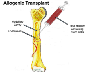
This type of transplant is a procedure that seeks to replace defective marrow cells with healthy cells..
The bone marrow harvesting technique is performed on the donor's pelvic bones, which are accessible for this procedure, although tissue from other bones can also be taken..
It consists of taking a sufficient amount of bone marrow from the iliac bones through large cannulas. The amount is calculated according to the weight of the recipient patient.

After a few weeks, through laboratory tests, it is determined if the patient's body accepted the transplant adequately and if his transplanted bone marrow is working.
Bone marrow transplantation is a complex procedure that can have complications. For this reason, a perfect study of both the donor and the recipient is required, as well as a specialized team of health professionals to guide them throughout the process..

Yet No Comments