Transcytosis characteristics, types, functions
The transcytosis it is the transport of materials from one side of the extracellular space to the other side. Although this phenomenon can occur in all cell types - including osteoclasts and neurons - it is characteristic of epithelia and endothelium..
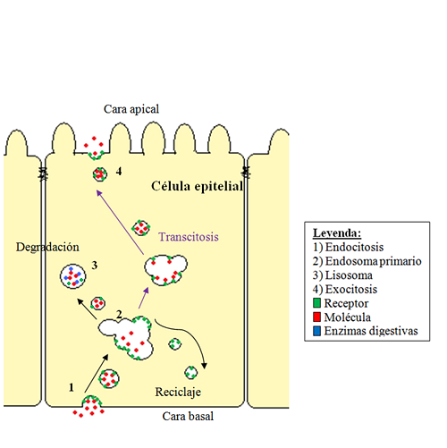
During transcytosis, the molecules are transported through endocytosis, mediated by some molecular receptor. The membranous vesicle migrates through the microtubule fibers that make up the cytoskeleton and on the opposite side of the epithelium, the contents of the vesicle are released by exocytosis.

In endothelial cells, transcytosis is an indispensable mechanism. Endotheliums tend to form impermeable barriers to macromolecules, such as proteins and nutrients.
Also, these molecules are too large to cross the transporters. Thanks to the transcytosis process, the transport of these particles is achieved.
Article index
- 1 Discovery
- 2 Process characteristics
- 3 Stages
- 4 Types of transcytosis
- 5 Functions
- 5.1 Transport of IgG
- 6 References
Discovery
The existence of transcytosis was postulated in the 1950s by Palade while studying the permeability of capillaries, where he describes a prominent population of vesicles. Later, this type of transport was discovered in blood vessels present in skeletal and cardiac muscle..
The term “transcytosis” was coined by Dr. N. Simionescu together with his working group, to describe the passage of molecules from the luminal face of the endothelial cells of the capillaries to the interstitial space in membranous vesicles.
Process characteristics
The movement of materials within the cell can follow different transcellular routes: movement through membrane transporters, through channels or pores, or through transcytosis.
This phenomenon is a combination of endocytosis, vesicle transport through cells, and exocytosis..
Endocytosis consists of the introduction of molecules into cells, encompassing them in an invagination from the cytoplasmic membrane. The vesicle formed is incorporated into the cytosol of the cell.
Exocytosis is the reverse process of endocytosis, where the cell excretes the products. During exocytosis, the vesicle membranes fuse with the plasma membrane and the contents are released into the extracellular environment. Both mechanisms are key in the transport of large molecules.
Transcytosis allows different molecules and particles to pass through the cytoplasm of a cell and pass from one extracellular region to another. For example, the passage of molecules through endothelial cells into circulating blood.
It is a process that needs energy - it is dependent on ATP - and involves the structures of the cytoskeleton, where actin microfilaments play a motor role and microtubules indicate the direction of movement..
Stages
Transcytosis is a strategy used by multicellular organisms for the selective movement of materials between two environments, without altering their composition.
This transport mechanism involves the following stages: first, the molecule binds to a specific receptor that can be found on the apical or basal surface of cells. This is followed by the endocytosis process through covered vesicles..
Third, intracellular transit of the vesicle occurs to the opposite surface from where it was internalized. The process ends with the exocytosis of the transported molecule.
Certain signals are capable of triggering transcytosis processes. A polymeric immunoglobulin receptor called pIg-R (polymeric immunoglobin receptor) undergoes transcytosis in polarized epithelial cells.
When the phosphorylation of an amino acid residue serine at position 664 of the cytoplasmic domain of pIg-R occurs, the process of transcytosis is induced.
In addition, there are proteins associated with transcytosis (TAP, transytosis-associated proteins) found in the membrane of the vesicles that participate in the process and intervene in the membrane fusion process. There are markers of this process and they are proteins of about 180 kD.
Types of transcytosis
There are two types of transcytosis, depending on the molecule involved in the process. One is clathrin, a protein molecule involved in the trafficking of vesicles within cells, and caveolin, an integral protein present in specific structures called caveolae..
The first type of transport, which involves clathrin, consists of a highly specific type of transport, because this protein has a high affinity for certain receptors that bind to ligands. The protein participates in the stabilization process of invagination produced by the membranous vesicle.
The second type of transport, mediated by the caveolin molecule, is essential in the transport of albumin, hormones and fatty acids. These vesicles formed are less specific than those of the previous group.
Features
Transcytosis allows the cellular mobilization of large molecules, mainly in the epithelial tissues, keeping the structure of the moving particle intact..
In addition, it constitutes the means by which infants manage to absorb antibodies from the mother's milk and are released into the extracellular fluid from the intestinal epithelium..
IgG transport
Immunoglobulin G, abbreviated, IgG, is a class of antibody produced in the presence of microorganisms, whether they are fungi, bacteria or viruses..
It is frequently found in body fluids, such as blood and cerebrospinal fluid. In addition, it is the only type of immunoglobulin capable of crossing the placenta..
The most studied example of transcytosis is the transport of IgG, from maternal milk in rodents, that cross the epithelium of the intestine in the offspring.
IgG manages to bind to Fc receptors located in the luminal portion of brush cells, the ligand receptor complex is endocyted in covered vesicular structures, they are transported through the cell and release occurs in the basal portion..
The lumen of the intestine has a pH of 6, so this pH level is optimal for the binding of the complex. Similarly, the pH for dissociation is 7.4, corresponding to the intercellular fluid on the basal side..
This difference in pH between both sides of the epithelial cells of the intestine makes it possible for immunoglobulins to reach the blood. In mammals, this same process makes it possible for antibodies to circulate from the yolk sac cells to the fetus..
References
- Gómez, J. E. (2009). Effects of resveratrol isomers on calcium and nitric oxide homeostasis in vascular cells. Santiago de Compostela University.
- Jiménez García, L. F. (2003). Cellular and molecular biology. Pearson Education of Mexico.
- Lodish, H. (2005). Cellular and molecular biology. Panamerican Medical Ed..
- Lowe, J. S. (2015). Stevens & Lowe Human Histology. Elsevier Brazil.
- Maillet, M. (2003). Cell biology: manual. Masson.
- Silverthorn, D. U. (2008). Human physiology. Panamerican Medical Ed..
- Tuma, P. L., & Hubbard, A. L. (2003). Transcytosis: crossing cellular barriers. Physiological reviews, 83(3), 871-932.
- Walker, L. I. (1998). Cell biology problems. University Publishing House.


Yet No Comments