The tracheostomy or tracheostomy is a surgical process that consists of making an incision in the anterior part of the neck, between the second and fourth tracheal rings, to open a direct airway between the trachea and the environment. A horizontal incision is made in an area called Jackson's safety triangle, two fingers above the suprasternal notch..
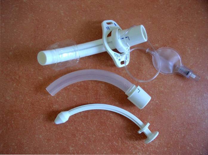
The resulting hole or stoma can serve as a direct airway or a tube called an endotracheal tube or tracheostome is placed through said hole, which allows air to enter the respiratory system without using the mouth or nose..
This procedure can be performed in a surgical room or on the patient's bed upon admission to the emergency department or intensive care department. It is one of the most widely used medical procedures in critically ill patients.
There are records and evidence of the use of the tracheostomy for more than 3,500 years by the ancient Egyptians, Babylonians and Greeks, to treat acute airway obstructions and thus save the lives of patients and animals.
Indications for a tracheostomy may be emergency or elective. In the first case, any acute situation that generates upper respiratory failure is included. In the second case, they are indicated for prolonged mechanical ventilation and the preoperative period of some major surgeries among others..
Among the most frequent complications are hemorrhages, tracheal stenoses, subcutaneous emphysema due to fistulas or loss of the airway, bronchospasm, serious infections of the airways and lungs, among others. These complications put the patient's life at risk.
Article index
Tracheostomies can be of various types and their classification can be made based on different criteria. Techniques, stoma location, and indications are the most commonly used criteria. In this sense, each of them are defined below..
The tracheostomy can then be:
Surgical tracheostomy is the classic tracheostomy performed under general anesthesia in an operating room. Percutaneous tracheostomy is performed on the patient's bed. Percutaneous tracheostomy currently tends to replace the classical surgical technique and has several technical modalities.
In turn, depending on the location of the stoma or tracheal opening, surgical and percutaneous tracheostomies can be:
According to their indication, tracheostomies can be divided into two types:
Elective tracheostomy It is indicated, for example, in patients with respiratory problems who are going to undergo major surgeries of the neck, head, chest or cardiac surgeries and who must remain intubated in the postoperative period for more than 48 hours.
Elective tracheostomy is also indicated before submitting the patient to laryngeal radiotherapy, in patients with degenerative diseases of the nervous system that can compromise the function of the respiratory pump, in some cases of comatose patients, etc..
Emergency tracheostomy It is used to solve emergency breathing problems that cannot be solved with endotracheal intubation and that are life-threatening. For example, patients with foreign bodies in the upper respiratory tract, mechanical obstructive problems due to neoplasms, etc..
The tracheostomy is placed permanently or temporarily. Permanents are generally used in patients who have undergone laryngotomies (removal of the larynx), usually for laryngeal cancer. The use of the tracheostomy, in most cases, is temporary and once the cause that indicates its use is resolved, the endotracheal tube is removed..
To avoid injury to organs adjacent to the trachea, both open and percutaneous surgical techniques are done within the Jackson triangle of safety. The Jackson safety triangle is an area that is shaped like an inverted triangle with the base up and the vertex down..
The anterior borders of the right and left sternocleidomastoid muscles form the sides of the triangle. The cricoid cartilage delimits the base of the triangle and the superior border of the sternal fork constitutes its vertex..
Because the percutaneous technique is quick, simple, easy to learn, and inexpensive, it has now been replacing the classical surgical technique. There are several modalities of percutaneous tracheostomy named after the physician who developed them..
The percutaneous wire-guided technique using progressive dilation was developed by Ciaglia. Later, this technique was modified by adding sharp, wire-guided forceps that allow for one-step dilation and was called the Griggs technique..
Later the Fantoni technique was developed. This technique uses a dilation that is performed from the inside of the windpipe outwards.
There are many other techniques that are nothing more than modifications of the original techniques, adding some instruments that increase the safety of the procedure, such as the concomitant use of a bronchoscope, among others. However, the most widely used techniques are those of Ciaglia and Griggs.
Although percutaneous tracheostomy is performed in the bed of the patient, it requires strict aseptic measures that include the use of sterile drapes and materials. Generally, two people should participate, the doctor who performs the procedure and an assistant.
Tracheostomy is indicated in any process that directly or indirectly affects the upper respiratory tract and generates respiratory distress that cannot be resolved via the laryngeal route. It is also indicated in prolonged connections to mechanical ventilation, such as airway after laryngotomies and in some preoperative of major surgeries.
The tracheostomy requires hygienic care and it is necessary to keep the cannula or tracheostome completely permeable so that it is free of secretions. The patient should avoid exposure to aerosols or other irritants or particles that are suspended in the air such as sand, earth, etc..
The main goal is to keep the pathway patent and avoid infection. When the tracheostomy is permanent, the patient must be trained in the care of the tracheostome and must attend a rehabilitation center to retrain speech.
Nursing care in hospitalized patients with tracheostomy has the same objectives. In these cases, the stoma should be disinfected at least once a day, ideally every eight hours. An antiseptic solution is used for this..
Once the stoma has healed, the endotracheal tube must be changed every four days, maintaining strict aseptic measures. The cannula must be aspirated to keep it patent. The patient must breathe in a humid environment to keep secretions fluid and facilitate their elimination.
The kit is prepared, which consists of a suction kit, gauze pads and sterile consumables, physiological and antiseptic solution, sterile gloves, a mask, a tape to hold the cannula and a bag to eliminate waste.
- It starts with hand washing
- An evaluation of the stroma is made, checking if there are reddened areas, edema or signs that suggest the presence of an infectious or hemorrhagic process.
- An aspiration of the trachea and pharynx is done following the technical procedure.
- The gauze is removed from the end of the cannula, washed with antiseptic solution, and a new gauze is placed. This gauze should not be cut to prevent the fibers that are shed from entering the trachea and causing abscesses or local infections..
- The cannula holding tape is changed. For this, sterile gloves, a mouth cover and glasses must be placed, and the help of a person with the same clothing must be available. This person should hold the end of the cannula while the tape is changed, avoiding the exit or expulsion of the tracheostome due to coughing or movements of the patient..
- Once this procedure is finished, the patient is put in bed and the pertinent annotations are made..
Tracheostomy complications are life-threatening. These can be acute while the patient has the endotracheal tube or in the process of placement, or they can appear later after the tracheostome has been removed..
The most frequent complications are hemorrhages, subcutaneous emphysema due to fistulas or loss of the airway, bronchospasm, serious infections of the airways and lungs. During the procedure, adjacent tissues such as the thyroid, vessels, or nerves may be injured..
As the tracheostome is removed and the trachea heals, stenosis may occur due to retractable scarring that tends to close the tracheal canal. This results in the need to re-enable a free airway and subject the patient to reconstructive surgery.
Tracheal stenosis is a very severe complication and the result of surgery has a high morbidity and mortality rate. However, percutaneous techniques have been associated with a lower frequency of complications compared to classical surgical techniques..


Yet No Comments