
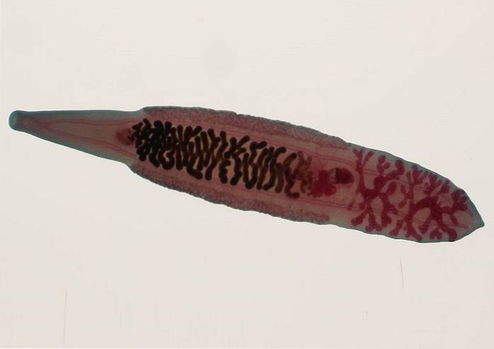
The trematodes they are a group of animals that belong to the phylum Platyhelminthes, specifically to the class Trematoda. They are flattened worms, with a typically leaf-shaped flattened body.
This class was first described in 1808 by the German zoologist Karl Rudolphi and is divided into two subclasses: Aspidogastrea and Digenea. Of these, the most studied and known is Digenea, since it includes flukes that cause certain pathologies in humans..
Diseases caused by flukes include bilharzia and schistosomiasis. They are related to the ingestion of contaminated water, as well as plants and animals contaminated with larvae of these parasites. This is why it is vitally important to maintain proper hygiene to avoid contagion..
Article index
Trematodes are considered multicellular eukaryotic organisms, because their cells have a cell nucleus that contains DNA in the form of chromosomes. They do not have a single type of cells, but they have a wide variety that each fulfill specific functions.
These animals are triblastic because during their embryonic development the three germ layers can be seen: endoderm, mesoderm and ectoderm. These undergo a process of differentiation to give rise to the tissues that make up the organs.
They are also cellophane. This means that they do not have an internal cavity known as a coelom. They are also protostome, so the mouth and anus are formed from an embryonic structure known as the blastopore..
They belong to the group of animals with bilateral symmetry, since they are made up of two equal halves.
Taking food into account, trematodes are heterotrophic organisms because they are not capable of synthesizing their nutrients, so they must feed on other living beings or substances made by them. Continuing with this, most are parasitic organisms, since they necessarily need to be inside a host to be able to subsist.
Almost all species are hermaphrodites and they contemplate, in their life cycle, the two types of reproduction that exist: asexual and sexual. Fertilization is internal, they are oviparous and have an indirect development.
The taxonomic classification of trematodes is as follows:
-Domain: Eukarya
-Animalia Kingdom
-Phylum: Platyhelminthes
-Class: Trematoda
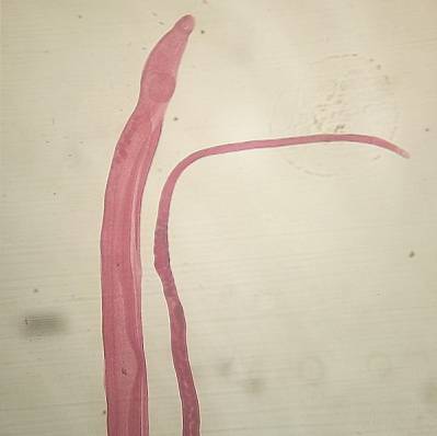
The organisms that belong to the Trematoda class are small in size. They measure about a few centimeters. This class is so wide that the morphology of the animals that make it up is quite varied. There are elongated, oval and flattened worms, among others.
In the place where the mouth opening is located, they have a suction cup, which helps this parasite to attach itself to its host. In addition, many of the trematode species have another sucker at the opposite end that is posterior.
The wall of the body of the trematodes is made up of several layers. From the outside to the inside, in order, they are described: an integument, which has no cilia and is quite thick; a layer of epithelial cells of the syncytial type; and finally, layers of muscle tissue, both circular and longitudinal.
Likewise, depending on the species, some may have certain structures on their body surface, such as spines. Orifices such as excretory and genital pores are also seen.
The digestive system of trematodes is incomplete. There is no anal orifice. It begins in the oral cavity, which continues with the pharynx and esophagus. The latter communicates with the intestine, which is divided into two tubes that are longitudinal. In these, the absorption of nutrients takes place.
It is protonephridial, made up of two ducts that are found on both sides of the body. Tubules that come from the so-called cells in flame flow into these ducts. In turn, they present a bladder that empties into an excretory pore.
It is quite simple. It is made up of several nerve cords, between which some communication is established through commissures. These cords have their point of origin in a plexus-type nerve conglomerate that is located in the cephalic part of the animal..
The vast majority of flukes are hermaphrodites. Due to this they present both female and male reproductive organs.
The male reproductive system is generally made up of a pair of testicles, from which the vas deferens arise, which end in the copulatory organ..
On the other hand, the female reproductive system consists of a single ovary, from which a duct (oviduct) arises that reaches the seminal vesicle. In addition to these structures, there is the uterus that is very close to the male pore.
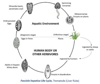
The life cycle of flukes is quite complex, since it involves a series of transformations until they reach adulthood. Likewise, this life cycle also includes the intervention of several intermediaries, which can be mollusks and crustaceans..
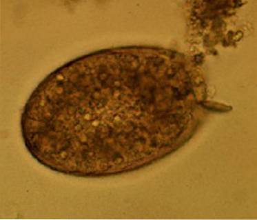
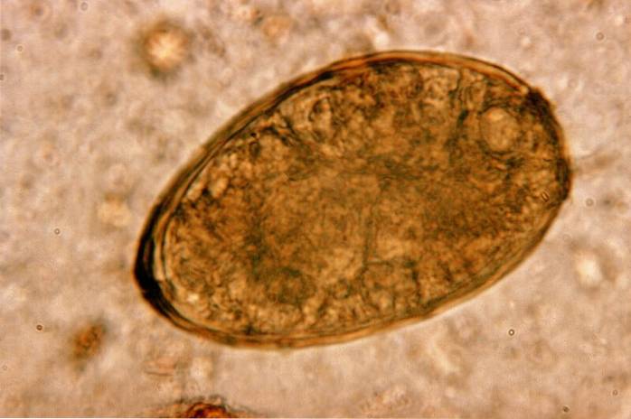
To explain the life cycle events of this parasite, the release of the eggs through feces or urine by the definitive host will be taken as a starting point..
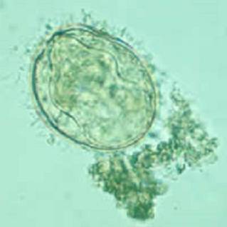
When the eggs are released from the host's body, either through feces or urine, they must reach an aqueous medium, since it requires certain humidity and temperature conditions to hatch..
When the egg is in the ideal conditions, a larva that is known by the name of miracidium forms inside it, which is generally surrounded by cilia, which facilitate movement and displacement through the aqueous medium..
A distinctive characteristic of this larva is that it does not have a mouth, which means that it has no way to feed. Due to this, this larva must move with the use of its cilia, until it finds a host before it runs out of nutrients..
Upon finding its ideal host, which is generally always a snail, the larvae penetrate its skin and enter its bloodstream. Within this host, the larva does not have a favorite organ to fixate and develop there. What you do take into account is the availability of nutrients.
Once the larva has settled in the snail's tissues, it undergoes another transformation, becoming the next phase: the sporocyst. This corresponds to a larva, which has the peculiarity of generating structures called germinative masses inside..
Immediately afterwards, the redias are formed, which constitute the next stage. These originate from each germ mass of the sporocyst. Redias already have a slightly more complex structure, with an easily identifiable pharynx and signs of the intestine and excretory system.
These break the sporocyst membrane and continue to develop inside the host (snail). It is important to highlight that several germinative masses (more than 40) begin to form on the wall of the redias, from which the next stage known as cercaria is formed. Of course, this happens when the temperature conditions are right..
Structurally speaking, the cercaria has the same internal structure as an adult trematode, with the exception that the reproductive system is not yet fully mature. They also have a tail that allows them to move freely through the middle.
However, the fence can be attached to a hard surface such as a plant and transformed into meta-fence. These can be passed on to a new host if the host ingests the plants. For example, if humans eat a plant that contains metacercariae, they travel through the digestive tract until they reach the duodenum..
In the duodenum they undergo a process of disembodiment and enter the bloodstream to initiate migration to other organs, such as the liver. There they fully mature and become adult parasites..
They can stay in the same place for long periods of time. There have even been cases of parasites that have lived there for up to several years.
Later the adults reproduce and begin to lay eggs, which are released mainly through feces..
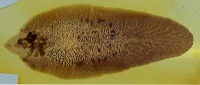
It is a species of trematode that belongs to the Digenea subclass. It is widely distributed throughout the world and is a parasite that affects some mammals, especially goats, cattle and sheep..
It is the causal agent of a disease known as fasciolosis. It is mainly housed in the bile duct, so the symptoms of infection by this parasite are centered in the liver, the most representative symptoms being pain in the right upper quadrant and disproportionate and painful growth of the liver..
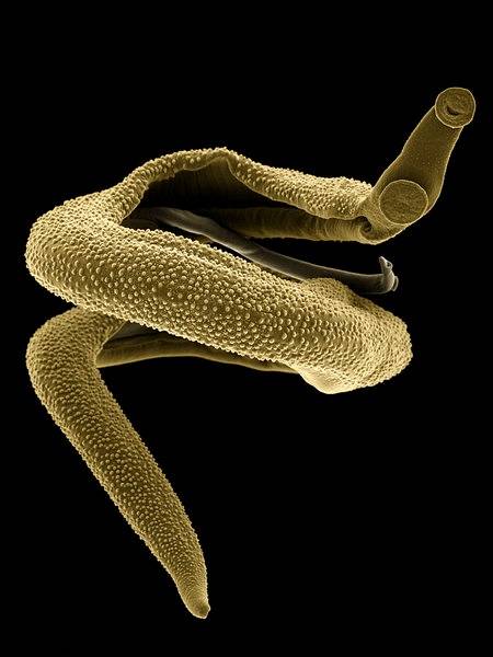
This is a parasite that belongs to the Digenea subclass. It is found mainly in developing countries such as Africans, some in Asia such as Yemen and others in South America such as Venezuela and Suriname.
Schistosoma mansoni It is a parasite of medical importance for humans, since it is responsible for a disease called Bilharziasis hepatic. The organs that are most affected by this parasite are the colon, rectum and, of course, the liver..
Although its natural hosts are other mammals such as cats, dogs, pigs and cows, it is also possible for man to become infected through contact with infected water.
It is an endemic parasite of the Mekong River basin in Cambodia. It is the cause of the highest percentage of cases of infection by Schistosoma in said region.
Schistosoma mekongi It causes serious damage to the body, as it feeds on the nutrients that circulate in the blood, as well as on red blood cells and blood proteins such as globulins. Of course, this has dire consequences for the host, since it stops perceiving the nutrients.
It is the largest trematode species that exists. It belongs to the order Echinostomida and can reach 75 mm in length. Morphologically it is very similar to Fasciola hepatica and has an estimated life time of approximately 6 months.
It can affect both man and pig. This parasite is known to cause a disease called fasciolopsosis, which is endemic to South Asian countries such as Indonesia, Vietnam, and Thailand..
This is an endemic parasite in some areas of Asia such as Indonesia, Korea, Japan and China, among others. It is the main responsible for the disease known as paragonimiasis. This affects several organs such as the liver, generating hepatomegaly, or the lungs, causing their function to be altered. It also causes coughing, diarrhea, and hives..
It is a parasite belonging to the Digenea subclass that is found mainly in Asian countries such as China, Japan and Taiwan. The most common way of transmission of this parasite is through the consumption of fish infected by the encyst larvae of the same.
These lodge in the bile ducts, where they reach adulthood, for which they present symptoms related to the liver such as painful hepatomegaly, jaundice and very high fever.
Contagion by a parasite belonging to the trematoda class has to do, in all cases, with the ingestion of one of its larval stages known as metacercariae. Depending on the trematode species, the infection vehicle is varied.
For some, such as those belonging to the genus Schistosoma, the contagion occurs by ingestion of water contaminated with the larvae of the parasite. On the other hand, in the trematodes of the genus Paragonimus, the contagion occurs by ingestion of river crabs, which constitute one of the hosts of the parasite.
In other genera, the consumption of fish that are infected by the larvae of the parasites is also involved..
Trematode infections cause complex symptoms that largely depend on the specific organ that is affected by the parasite..
Since most parasites lodge in the digestive tract, the most common symptoms have to do with them. In this sense, the most representative intestinal symptoms of trematode infection are the following:
- Abdominal pain, especially in the right upper quadrant
- Jaundice
- Exaggerated increase in liver size
- Biliary colic
- Repetitive belching
- Diarrhea
Likewise, when the affected organs are others, such as the lung, the central nervous system, the skin or the bladder, the symptoms are:
- Frequent urinary infections
- Burning when urinating
- The urge to urinate very often
- Intense itching
- Chronic cough, which may be accompanied by bloody sputum.
- Dyspnea or shortness of breath.
- Seizures
- Muscular weakness
- Paralysis, which can be temporary or permanent.
The diagnosis of infections caused by trematodes is simple, since the doctor, knowing the symptoms manifested by the patient, can guide his diagnosis towards an intestinal parasitosis. In such a way that the tests that are carried out are only to establish a differential diagnosis. The most used exams are the following:
This is the test most often used to specifically diagnose intestinal parasite infections. Since most of these release their eggs using feces as a vehicle, examining them determines the presence of the eggs and therefore demonstrates the infection.
In this test the stool is examined at the microscopic level and a histological study is performed. It is a non-invasive examination and, generally, quite accessible from the economic point of view.
For patients with pulmonary symptoms, the doctor may collect a sample of the sputum and send it to a laboratory to examine it for eggs..
This test also has high reliability, although it is used less frequently, since most patients present with digestive symptoms..
Through a simple blood test, it is possible to identify antibodies against this parasite. This type of test is also effective, although the stool test is generally the most common..
Through examinations such as X-rays, ultrasound or a computerized axial tomography, lesions in some internal organs can be evidenced. These tests are not used for a diagnosis, but rather in a complementary way to assess the extent of damage caused by the parasite..
Because flukes are parasites, the main treatment option is anthelmintic drugs. The most commonly prescribed are albendazole and praziquantel. These drugs have a harmful effect on the parasite, intervening in its metabolism, eventually causing its death..
Medications that alleviate the symptoms caused by the parasite may also be prescribed, such as pain relievers and anti-inflammatories, among others..


Yet No Comments