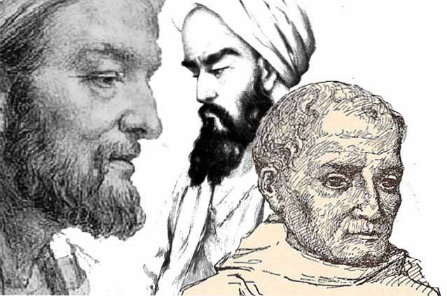
Trichomonas hominis it is a multiflagellate protozoan that lives as a commensal in the intestinal tract of some mammals. It was first observed and described by the English physician Casimir Devaine in 1854. It is also known as Pentatrichomonas hominis because in its structure it has five flagella.
It is considered a non-pathogenic organism for humans since, despite being found with certain frequency in the intestine of the human being, on very few occasions it causes damage and stimulates the appearance of symptoms. However, it is more common to find it in populations settled in warm places and within these, in children under 10 years of age.
Article index
The taxonomic classification of Trichomonas hominis is the next:
Trichomonas hominis it is a unicellular organism, which means that it is made up of a single cell. That cell is of the eukaryotic type. This implies that its genetic material is delimited by a membrane, enclosed in a cellular organelle known as the nucleus..
Being a parasite, this protozoan necessarily needs another living being in order to survive. In this case, it lodges in the intestinal tract of some mammals and benefits from the products of their digestion..
Despite this, it could almost be said that he lives under a commensal relationship, since there are very few occasions in which he triggers a pathological reaction.
Trichomonas hominis it lives in the large intestine of some mammals such as man and some rodents. The area of the large intestine where this protozoan tends to be located is in the cecal area.
From a geographical point of view, the protozoan is abundant in places with a warm climate.
The trichomonas hominis it is a heterotrophic organism. It feeds on the substances that circulate through the digestive tract of mammals that it parasitizes..
Feeding is carried out through phagocytosis. Through this process, the protozoan surrounds the food particles with its plasma membrane and incorporates them into its cytoplasm so that they are processed by the digestive enzymes that are inside the protozoan..
In this type of protozoa, the reproduction that is observed is asexual, it does not require the union of gametes.
The process by which it reproduces Trichomonas hominis is the longitudinal binary fission. In this, the duplication of the protozoan DNA occurs. Subsequently, each copy goes to one end of the cell and it begins to lengthen.
Finally, the cytoplasm undergoes a strangulation along the longitudinal axis, until the cell divides completely, originating two cells genetically identical to the progenitor..
The protozoan Trichomonas hominis It only has one life form in its life cycle, the trophozoite< es decir, no presenta quistes.
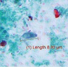
The trophozoite has a shape similar to that of a pear. It has approximate measurements of between 5-15 microns, although some have been recorded that have reached up to 20 microns. Likewise, it is a cell with a single nucleus, which is located towards the anterior pole of the cell..
The nucleus is associated with an endosome; a set of vesicles that have been generated through endocytosis that contain material that has been captured outside the cell.
Seen under the microscope, it can be seen that it has a total of five flagella, of which one is found on the cell surface, forming a kind of undulating membrane. The rest of the flagella are arranged oriented towards the anterior pole.
They have a structure known as axostyle, a set of microtubules that are very close together. These run through the entire axis of the cell and can even extend beyond this.
These microtubules are surrounded by a sheet that forms a tube that may or may not be hollow. This structure has a function in relation to locomotion.
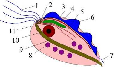
Likewise, there are structures known as blepharoplasts, basal corpuscles from which flagella originate..
In its cytoplasm it does not present mitochondria, but a Golgi apparatus, which is called the parabasal body.
This protozoan has several possible hosts, all mammals: rodents, dogs, and primates, like man. However, flies can sometimes act as indirect vectors, as they frequently carry fecal remains on their extremities..
The site of the human body where this protozoan is found is the large intestine, mainly the cecum. There it feeds on the intestinal contents. It is always in the trophozoite state, since it does not present cysts.
The trophozoites are released through the feces. They can be ingested by a new host when he ingests food or water contaminated with fecal particles infested with trophozoites of Trichomonas hominis.
Once inside the organism of the new host, the trophozoites are transported through the digestive tract to the large intestine, finding their ideal habitat. There they begin to reproduce and spread through the large intestine, although their site of predilection is the cecum..
Later they are expelled with the feces so that the cycle continues.
The Trichomonas hominis it is a protozoan that generally does not cause any pathology. However, when for some reason it begins to reproduce in an uncontrolled way, considerably increasing its number in the intestine with the consequent irritation of the intestinal mucosa.
The main transmission mechanism of the Trichomonas hominis It is through the ingestion of food and water contaminated with stools with trophozoites.
An individual may be found to be infected with Trichomonas hominis without presenting any type of symptoms. This is what happens most frequently, since this is a non-pathogenic protozoan for humans.
Despite this, when the number of parasites is very abundant, they tend to erode and inflame the intestinal mucosa, with the consequent diarrhea-like symptoms:
The main way to diagnose infection by Trichomonas hominis it is through the study of fresh feces. Once the sample is obtained, it is observed under the microscope to identify the presence of protozoan trophozoites..
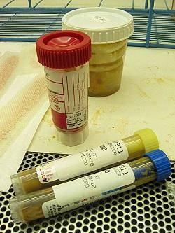
Likewise, there are other diagnostic methods, among which the stool test or stool culture stands out. In this, a culture with stool samples is carried out in order to detect any microorganisms that grow there.
The finding of Trichomonas hominis in the stool may be accidental in some routine examination. Doctors choose not to prescribe any treatment if the individual does not present any symptoms.
Now, if your finding is linked to the persistence of any intestinal symptoms such as diarrhea or colic, it is necessary to use some medicine.
In this case, the drugs to treat intestinal parasites are almost always the same. Among the most used is metronidazole, an antiparasitic whose mechanism of action focuses on nucleic acids, inhibiting their synthesis and therefore, preventing the multiplication of protozoa.
Other treatment options are tinidazole, secnidazole, and ornidazole..


Yet No Comments