The scarpa triangle, Also known as the femoral triangle, it is a triangular anatomical area, with a lower vertex, located in the antero-superior part of the thigh. The way to expose the femoral triangle and properly identify its limits is by placing the patient's thigh in flexion, with a slight lateral rotation.
The inguinal ligament forms the base of this area, and the sartorius and adductor longus muscles of the leg, its sides. It is a region that acquires great importance in topographic anatomy, since it contains the main blood vessels of the lower limb, the femoral artery and vein, as well as the primordial neurological branch and the femoral nerve. The Scarpa triangle is the most accessible region to identify these structures.
The femoral artery is the main feeding vessel in the lower limb, and through it, other important arteries in the body can be accessed for complex surgical procedures. This technique is used in the specialty known as interventional radiology and in the subspecialty of cardiology called hemodynamics..
In emergency medicine, the health professional must be aware of this area, since if a traumatized patient has significant bleeding in the leg, difficult to control and threatening his life, it can be stopped by blocking the femoral artery from the Scarpa triangle.
The obstruction of the femoral artery by means of a tourniquet in case of trauma, is a procedure that can save the patient's life.
Article index
The lower limbs begin their formation around 4ta gestation week. As the legs are formed, the differentiation of the other structures also begins.
For the 10ma week, all the elements are completely differentiated, including the blood vessels, the nerves and the skin. The area that is recognized as femoral triangle also completes its training with the differentiation of the inguinal ligament.
The groin is the region of the body that joins the abdomen with the lower limbs. In its cutaneous projection, it is the oblique area that is located towards the medial plane, just below the trunk, at the hip joint, and that joins the lower abdomen with the lower limbs..
However, deeply the inguinal region encompasses a wider area extending from the lower insertion of the abdominal muscles to the inguinal ligament..
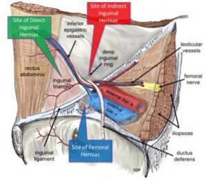
The inguinal or Poupart's ligament extends from the antero-superior prominence of the ilium to the symphysis pubis. Forms the lower border of the inguinal region and the upper border of the anterior femoral region.
This ligament is the anatomical landmark that delimits and separates the inguinal from the femoral region. Knowing its location is essential for the description of some pathologies and for the performance of clinical and surgical procedures..
Within the inguinal region is the inguinal canal, which contains the spermatic cord in men and the round ligament of the uterus in women. The path through the inguinal canal is an area of weakness in the abdominal wall where inguinal hernias frequently occur..
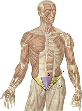
The femoral region is located just below the inguinal region. In its anterior part, the femoral or Scarpa triangle is described, which is an anatomical division that is used to facilitate the study of this area..
The femoral triangle is an area that is located in the anterior and upper part of the lower limb. Its superficial projection is exactly in the groin.
This anatomical division is located below the inguinal region. It is shaped like an inverted triangle, its vertex is at the bottom and its base is at the top..
It is bounded above by the inguinal or Poupart ligament, laterally by the sartorius muscle and medially by the adductor longus muscle. Its vertex is formed by the intersection of these two muscles.
Covering this entire area is a fibrous and elastic structure called cribriform fascia, which is an extension of the transverse fascia that comes from the abdomen. This tissue covers the blood and lymphatic vessels found in the femoral region, up to 4 cm below the inguinal ligament..
Within the limits of the femoral triangle are the femoral artery, vein, nerve, and lymph nodes.
The femoral artery is the main feeding vessel of the lower limb. It is the continuation of the external iliac artery, a branch of the common iliac artery that is a direct branch of the aorta. It is a large-caliber blood vessel that is responsible for guaranteeing the blood supply to all the muscles in the region..
For its part, the femoral vein is the main route of blood return from the lower limb.
The femoral nerve is an important structure that provides mobility and sensitivity to the leg and foot, and the femoral lymphatic vessels communicate the superficial with the deep system and have an important lymph node station in the groin.
The femoral triangle is the region in which these structures are most superficial, so it is easy to identify them on physical examination if the anatomical limits of the area are known..
The femoral triangle contains structures that are essential for the function of the lower limbs. Knowing the location of this region guarantees safe access to these anatomical elements, and it is also the only way to carry out an exploration appropriate to the physical examination..
The femoral artery is easily palpable at this level. When the patient's peripheral pulses are weak, this is one of the arteries in which the heart rate can be verified on physical examination..
It is also an accessible route when laboratory tests with arterial blood are needed specifically..
The femoral vein is also used when catheterization of common venous lines or for taking laboratory samples is not possible..
In procedures such as neurological block for lower limb surgeries, the femoral triangle is used as a reference to find the femoral nerve and be able to practice this technique safely.
In addition, it is an area in which the lymph nodes are usually examined as it provides information about the status of the entire lower limb. Inflammation of these nodes can indicate the presence of any infectious process but it can also be a sign that a malignant disease, such as melanoma, is metastasizing lymph nodes.
In the case of polytraumatized patients, the femoral area is highlighted as an important point when stopping profuse bleeding from the lower limb that threatens the patient's life.
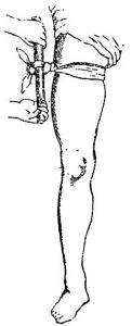
By making a strong tourniquet in this area, it is possible to obstruct the flow of blood through the femoral artery, which avoids the massive loss that can cause death..
In any surgical procedure of the inguinal or femoral region, it is important to know all the anatomical landmarks that delimit these areas as well as the location of the structures they contain.
In the case of inguinal hernia or femoral hernia repair surgery, the procedure involves reinforcing the entire area with a material that is sutured to the inguinal ligament and cribriform fascia.
The surgeon must be familiar with the area to avoid injuring any of the structures contained in these regions, since they are the ones that guarantee the correct functioning of the lower limb..
Lymph nodes located in the femoral triangle are a frequent location for metastases due to malignant tumors of the lower limbs. When they are inflamed, surgical procedures must be performed for their study and treatment..
The inguino-femoral lymph node dissection It is a surgery in which all the fat is removed with lymph nodes that are located in the inguinal and femoral regions.
All this lymphatic tissue is attached to the blood vessels and femoral nerves, therefore, when performing this procedure, the location of the vascular and neurological structures must be taken into account in order to extract the necessary material without leaving sequelae in the patient..
Both interventional radiology and hemodynamics are subspecialties of radiology and cardiology respectively, which are responsible for diagnosing and treating diseases of the blood vessels.
Through long guides of surgical material, arteries and veins are canalized, special contrast is injected and X-rays are taken that allow drawing the vascular map of the patient and observing the problem that he presents..
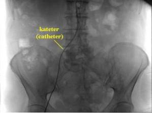
The most commonly used routes to perform these procedures are the femoral vessels. Right at the level of the femoral triangle, the vessel to be studied is identified, either the artery or the vein, and a special catheter is inserted. These procedures are known as angiography.
The femoral blood vessels continue with the great vessels of the abdomen, the aorta, and the vena cava, which open directly into the heart. For this reason, through the location of the femoral route, the catheter is directed to where it is required to inject the contrast and diagnose and treat the pathology..
For example, when a patient presents an obstruction in an artery of the heart by a blood clot and this causes a myocardial infarction, the point of the obstruction can be found through the passage through the femoral artery..
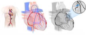
Once the desired point in the heart is reached, the severity of the problem can be observed by taking X-rays or radiological video (fluoroscopy) and injecting an agent that dilutes the clot to prevent damage to the heart muscle..

Yet No Comments