
The trophoblast it is one of the tissues present in the embryos of mammalian animals; is fundamental to the processes known as implantation Y placentation that take place, in humans, during the first weeks of gestation.
The embryonic development of mammals begins with fertilization, that is, with the formation of the zygote, which occurs after the cellular and nuclear fusion of a mature egg and sperm. These are the sex cells or haploid gametes from a male and a female individual..
About 30 hours after fertilization, the new cell that forms begins to divide by mitosis, in a process known as cleavage zygote.
This implies a rapid increase in the number of cells, now known as blastomeres and occurs as the zygote makes its way through the fallopian tubes to the uterus and from the ovary, where the eggs reside.
When the zygote reaches a 9-cell stage, the blastomeres undergo a metamorphosis and line up in an orderly fashion, forming a compact ball, where two groups of cells will then differentiate, some internal and others external..
Three days after fertilization, by which time the embryo has only about 32 cells, the stage known as morula, which is the one that enters the uterine cavity. In the morula, a fluid-filled cavity begins to form that derives from the uterine cavity..
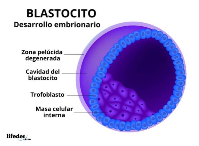
The fluid in the cavity (called blastocyst cavity) it separates the cells into two well-defined layers: an internal one, located centrally, and an external one that covers it; this stage is called blastocyst. The inner cell layer or mass is called embryoblast and the outer layer trophoblast.
The embryo's own structures will later form from the embryoblast as embryonic development progresses, while the embryonic portion of the placenta is derived from the trophoblast, which performs nutritional and protective functions for the embryo..
Trophoblast cells, during the first 10 days of pregnancy in humans, secrete a protein known as "early pregnancy factor", which enters the woman's blood circulation approximately 48 hours after fertilization and is the basis for rapid tests. of pregnancy.
The prefix "trophus”In the term trophoblast, it alludes to the Greek word used for nutrition or feeding, since the main function of this embryonic tissue is to participate in the formation and development of the tissues that will feed the embryo during its stay in the uterine cavity (the placenta).
The participation of the trophoblast in the embryo is crucial for its success, so to speak, since this thin layer of cells plays a fundamental role in the processes known as implantation and placentation..
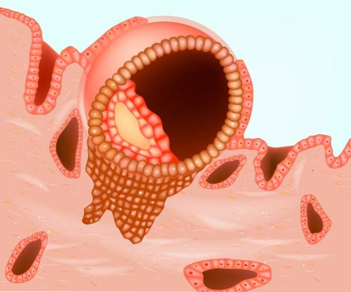
Implantation refers to the anchoring or "invasion" of the embryo to the uterine walls, from where it will later be fed by the maternal tissues, once the placenta is formed, which will also protect it..
Implantation occurs as a consequence of efficient temporal synchronization, cell adhesion and migration processes and adequate communication between the embryo and its mother through different molecular and hormonal factors, many of them derived from trophoblast cells..
In this process are involved, among other factors, the transforming growth factor-β, that regulates the proliferation and "invasion" of trophoblast cells into maternal tissue through their interaction with a series of membrane receptors.
The placenta is the tissue that allows the exchange of information between the embryo and its mother, as well as the nutrition and support of the embryo during its development; this tissue is essential for fetal health and for the successful completion of pregnancy and is derived from extra-embryonic tissues.
The trophoblast has a dual function in placentation and implantation, since it is responsible for, as the embryo implants, forms the placenta.
The trophoblast, as the embryo attaches to the endometrial epithelium (implants), proliferates and separates into two layers:
The syncytiotrophoblast consists of a layer formed by a syncytium, that is, it is a layer of cells that do not have divisions or membranes that separate them from each other, resulting in a large multinucleated cytoplasmic “mass”.
This layer, which derives from the fusion of the cells present in the cytotrophoblast, achieves infiltrate in the epithelium of the endometrium, which is the inner layer of the uterus, fixing the embryo to the maternal tissue; this occurs thanks to the endometrial cells undergoing programmed cell death, facilitating infiltration.
The syncytiotrophoblast produces a hormone known as human chorionic gonadotropin (known as hCG for its acronym in English), which is responsible for maintaining hormonal activity in the ovaries during pregnancy.
The cytotrophoblast, instead, it is the layer closest to the embryoblast and consists of a series of ovoid-looking cells, with a single nucleus.
As embryonic development progresses, irregular spaces called lagoons, which are initially filled with uterine fluids and secretions.
Meanwhile, this layer of the trophoblast grows even further towards the uterine wall and perforates the walls of the blood vessels present there, allowing the blood to flow towards the gaps, in order to support the development of cells in the cytotrophoblast.
The cytotrophoblast also proliferates during implantation, in the periphery of the placental tissue, until it joins it and allows the formation of vasculature that communicates the embryo with the mother..
A very common pathological condition that is related to the trophoblast is Gestational trophoblastic disease, that has to do with its proliferation in pregnant women, the most common consequence of which is spontaneous abortion or fetal death.
It is generally defined as a group of tumors, that is, groups of cells that divide uncontrollably, that derive from the trophoblast and that surround or cover the blastocyst (the developing embryo), forming between the sites comprised by the amnion and the chorion..
The risk factors associated with these conditions are age (too young or over 35 years old), the amount of hormones produced during pregnancy, the presence of other tumors in the uterus, high blood pressure, etc..
Although these diseases are rare, two different types have been described:
Molar pregnancy is the most common type and is generally associated with defects in gametogenesis or during fertilization. It refers to a group of tumors that look like fluid-filled sacs that grow slowly..
Choriocarcinoma is the malignant form of this disease, as it spreads rapidly to the muscular layer of the uterus and even to other regions of the body..


Yet No Comments