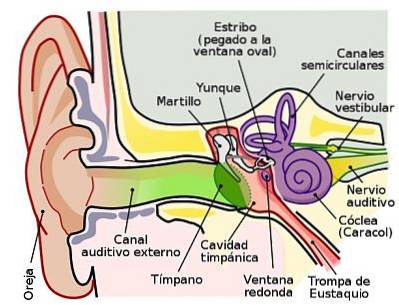
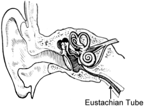
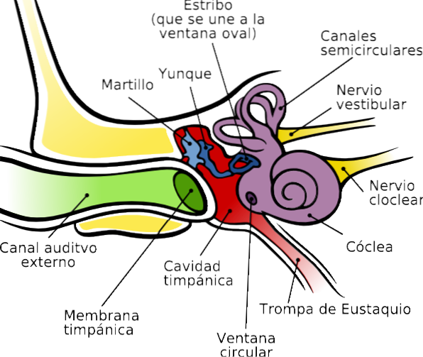
The eustachian tubes are two canals, right and left, each associated with the tympanic cavity of the middle ear on the corresponding side (right and left) and that communicate, respectively, said compartments of the auditory system with the nasopharynx.
It is usually called the "Eustachian tube" in honor of the anatomist who discovered it in the 16th century, but it is also commonly called "tuba", "auditory tube", "tympanic tube" or "pharyngotympanic tube".
These names refer to the relationship of such structures with the auditory system and more strictly with the tympanic cavity..
The Eustachian tube has no direct function in the processes of mechanical transmission of sound waves, nor in those of sensory processing or nerve conduction inherent to auditory function. However, by allowing the equalization of the pressures on both sides of the tympanic membrane, it contributes to its having the appropriate degree of tension for the faithful transmission of these waves.
- The Eustachian tube apparently develops from an embryonic structure known as the “tubotympanic recess”, which probably originates, in turn, in the vicinity of the first embryonic pharyngeal pouch.
- It is a duct between 35 and 45 mm in length.
- It leads, following a downward trajectory, forward and inward, from the tympanic cavity in the middle ear to the nasopharynx, a space located behind the nasal passages, in continuity with them and above the soft palate.
Taking into account the beginning of its journey from the tympanic cavity and its end at the level of the nasopharynx, the Eustachian tube can be considered divided into:
- an initial journey or bone portion Y
- a final segment or cartilaginous portion, both united in a narrow (stenosed) region called the isthmus.
It corresponds to the first third of the length of the Eustachian tube; is a cylindrical and anterior prolongation of the tympanic cavity.
It occupies a kind of semi-channel in the peñasco of the temporal bone and can be considered a portion of the pneumatic (air-filled) area of said bone, together with the tympanic cavity itself and the air cells of the mastoid process..
It is related cranially (above) with the semichannel for the tensor chorda tympani; in front and outside with the tympanic portion of the temporal bone, and behind and inside with the carotid duct.
It is represented by the lower or distal two thirds of said tube, once it leaves the thickness of the temporal rock.
This portion is considered to be a diverticulum of the pharynx and is found on the underside of the skull base, in a groove between the greater wing of the sphenoid (a bone at the base of the skull) and the petrous portion of the temporal bone..
The structure of its wall is made of cartilage of the elastic type, and it is a lamina completed at the end, caudally, by connective tissue.
It is externally related to the tensor veli palate, the inferior maxillary nerve, and the middle meningeal artery; inside, with the levator veil palate and the pharyngeal recess.
It is the hole that marks the mouth of the tube in the nasopharynx. There are two, one on each side and for each horn.
Through these holes, and accessing them through the external nostrils, the catheterization of the tubes can be practiced during certain surgical procedures.
This fact makes it important to know the location of said hole, which is located on each side on the corresponding external wall of the nasopharynx and approximately between 1 and 1.5 cm:
Both the tympanic cavity and the Eustachian tube are internally lined by a mucous epithelium that has certain differential characteristics depending on the segment in question..
The bone portion is covered, like the tympanic cavity, by a kind of "mucoperiosteum" that is normally characterized by an epithelium of cuboidal cells, flattened and without cilia..
The mucosa of the cartilaginous portion, for its part, is more similar to the pseudostratified respiratory epithelium of the nasopharynx, with columnar and ciliated cells.
The functions of the Eustachian tube are related to its character as a conduit that communicates the tympanic cage with the nasopharynx and that allows the passage of liquid and / or air flows between both cavities.
It should be noted that the periosteal mucosa of the middle ear tympanic cavity is continuously producing mucous secretions that are drained into the nasopharynx through these tubes..
This drainage is facilitated by the action of gravity, since these tubes follow an inclined and descending path and the exit orifice in the nasopharynx is at a lower level than that of entry in the eardrum..
Added to this is the movement of the cilia of the epithelium of the cartilaginous portion that actively contributes to pushing said mucus downward..
The tubes communicate the tympanic cavity with the gas contained in the nasopharynx, which in turn is in pressure equilibrium with atmospheric air.
Hence, when the tubes are open, the pressure of the gas in the tympanic cavities is the same as the pressure of atmospheric gas..
This equilibrium in pressure is given by air flow in one direction or another. When the atmospheric pressure is low with respect to the tympanic, the gas moves outwards and the tympanic pressure also drops.
On the contrary, when tympanic pressure drops, gas flows from outside and tympanic pressure rises..
The result of this equilibrium makes the pressure that the atmosphere exerts on the face of the tympanic membrane that faces the external auditory canal, is exactly the same as the pressure that that same atmosphere exerts on the face of the membrane that faces the tympanic cavity.
This pressure balance between both faces of the tympanic membrane is a fundamental condition for the latter to have the appropriate shape and degree of tension that allow optimal transmission of sound vibrations..
The cartilaginous portion of the tubes is collapsed, that is, the tubes are closed and there is no communication between their ends.
When the phenomenon of swallowing occurs, the tubes open, either passively or by the action of the tensor veli palate muscle..
Swallowing is a process that occurs intermittently and at more or less short intervals, since mucous secretions are continuously produced along the pharynx and saliva at the level of the oral cavity, secretions that are ingested by this frequent swallowing..
Some alterations in the function of the Eustachian tube are related to its obstruction and the rupture of the pressure balance between the external auditory canal and the middle ear, which leads to a considerable reduction in the efficiency of the transmission of sound waves and the production of a certain degree of deafness.
When reaching considerable heights, such as when ascending in an airplane or climbing a mountain, the atmospheric pressure drops and the air contained in the tympanic cavity expands and rejects the tympanic membrane outwards..
If no swallowing movements are made, the higher internal pressure can suddenly open the tubes producing a “snap”..
When altitude is lost, reverse pressure changes occur. That of the eardrum becomes lower than the atmospheric one, which produces a retraction or puckering of the membrane with the production of deafness.
In this case, the spontaneous opening of the tubes will not occur, which tend rather to collapse..
To correct the difference, it is mandatory to perform maneuvers such as forced swallowing, yawning or the Valsalva maneuver.
A complication that can occur, apart from the production of pain, is the rupture of the tympanic membrane. Phenomenon that does not usually occur unless the pressure difference exceeds between 100 and 500 mm Hg, which usually happens to divers.
Apart from the circumstantial changes of the surrounding pressure, various pathologies can lead to the obstruction of the tubes.
These include the common cold and other upper respiratory infections, chronic middle ear infections, rhinitis, hypertrophy of the adenoids and alterations of the nasal septum..

Yet No Comments