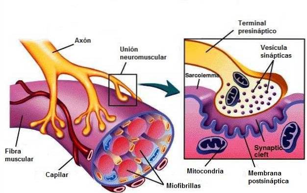
The neuromuscular junction or neuromuscular plate is the synapse between a motor neuron and a muscle. Thanks to the transmitted impulses, the muscle can contract or relax. Specifically, it is the connection between the terminal button of a neuron and the membrane of a muscle fiber..
The terminal buttons of neurons connect to the motor terminal plates. The latter refer to the membrane that receives nerve impulses from a neuromuscular junction..
This type of synapse is the most studied and the easiest to understand. To control a skeletal muscle, a motor neuron (motor neuron) synapses with a skeletal muscle cell..
Article index
The neuromuscular junction is made up of the following elements:
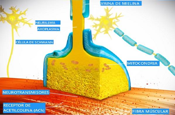
This neuron is called presynaptic because it emits nerve impulses or action potentials. Specifically, nerve impulses travel through the axon of this neuron to the terminal button that is located very close to the muscle. This termination has an oval shape about 32 microns wide..
In the terminal button are the mitochondria and other elements that allow the creation and storage of acetylcholine. Acetylcholine is the main neurotransmitter for muscle stimulation.
Many authors refer to this element as an alpha motor neuron, as it is a type of neuron whose axon synapses with extrafusal muscle fibers from a skeletal muscle. When activated, it releases acetylcholine, causing muscle fibers to contract..
The terminal button of the neuron and the muscle membrane are not in direct contact, there is a small space between them.
It is made up of one or more muscle cells. These target cells constitute a muscle fiber.
There are different types of muscle fibers. The muscle fibers that are innervated at the neuromuscular junction are called extrafusal muscle fibers. They are controlled by alpha motor neurons and are responsible for the force that arises from the contraction of a skeletal muscle.
Unlike these, there are other types of muscle fibers that detect the stretching of a muscle and are parallel to the extrafusal fibers. These are called intrafusal muscle fibers..
A muscle fiber is made up of a bundle of myofibrils. Each myofibril is made up of overlapping filaments of actin and myosin, which are responsible for muscle contractions..
Actin and myosin are proteins that form the physiological basis of muscle contraction..
Myosin filaments have small protrusions called myosin cross-linking bridges. They are the intermediaries between myosin and actin filaments and are the mobile elements that produce muscle contractions..
The parts where actin and myosin filaments overlap are seen as dark bands or streaks. For this reason, skeletal muscles are often called striated muscles..
Myosin cross-linking bridges "row" along the actin filaments so that the muscle fiber shortens, contracting.
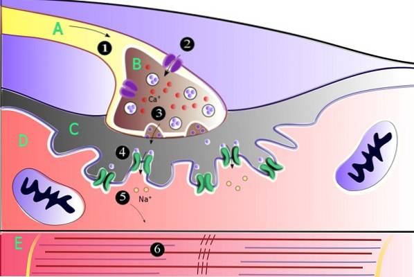
Neuromuscular junctions are located in the grooves across the surface of the muscle fibers. When an action potential or electrical impulse travels through the neuron, its terminal button releases a neurotransmitter called acetylcholine..
When a certain amount of acetylcholine accumulates, it produces the so-called end plate potential in which the muscle membrane depolarizes. This potential is much broader compared to that produced between two neurons..
The terminal binding potential always gives rise to the activation of the muscle fiber, expanding this potential throughout the entire fiber. This causes a contraction or jerk of the muscle fiber..
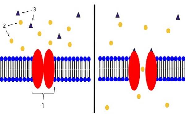
Depolarization is the reduction of the membrane potential of a cell. When a muscle fiber is depolarized, calcium channels begin to open, allowing calcium ions to penetrate into them. This phenomenon is what causes muscle contraction.
This is because calcium works as a cofactor, which helps the myofibrils extract energy from the ATP that is in the cytoplasm..
A single nerve impulse from a motor neuron results in a single contraction of a muscle fiber. The physical effects of these shocks are much longer than those of an action potential between two neurons.
This is due to the elasticity of the muscle and the time it takes to rid the cells of calcium. In addition, the physical effects of a set of nerve impulses can accumulate leading to a prolonged contraction of the muscle fiber..
Muscle contraction is not an all or nothing phenomenon, as are the contractions of the muscle fibers that make up the muscle. On the contrary, the force of the shock is determined by the average discharge frequency of the different motor units..
If at a given moment they discharge many motor units, the contraction will be more energetic, and if they discharge few, it will be weak.
The pathologies of the neuromuscular junction can affect the terminal button of the motor neuron, or the membrane of the muscle fibers. For example, botulism produces an alteration and inhibition in the release of acetylcholine, both in the skeletal muscles and in the autonomic nervous system.
It is acquired by consuming contaminated food, mainly. Within a few hours it produces a progressive and rapid muscle weakness.
On the other hand, myasthenia gravis, which is the best known neuromuscular disease, appears due to the inflammation of acetylcholine receptors. It arises from antibodies that these patients have that attack these receptors.
Its main symptom is weakness of the voluntary skeletal muscles. It is observed mainly in the muscles that participate in the breathing, salivation and swallowing; as well as on the eyelids.
Another example of pathology of the neuromuscular junction is Lambert-Eaton syndrome, which consists of an autoimmune disease in which the immune system mistakenly attacks the calcium channels of motor neurons.
This generates an alteration in the release of acetylcholine. Specifically, the propagation of the motor action potential is blocked. Muscle weakness is also observed, in addition to tumors.

Yet No Comments