The cell junctions They are the contact bridges that exist between the cytoplasmic membranes between adjacent cells or between a cell and the matrix. The junctions depend on the type of tissue studied, highlighting the existing connections between epithelial cells, muscle cells and nerve cells..
In cells there are molecules related to adhesion between them. However, additional elements are needed to increase the stability of the bond in tissues. This is achieved with cell junctions.
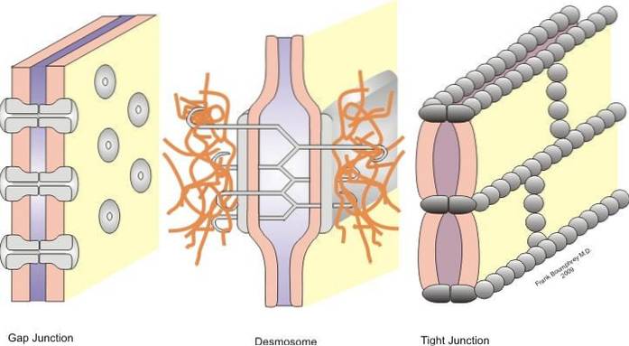
Junctions are classified into symmetric junctions (tight junctions, belt desmosomes, and slit junctions) and asymmetric junctions (hemidesmosomes).
Tight junctions, belt desmosomes, point desmosomes, and hemidesmosomes are junctions that allow anchoring; while the cleft junctions behave as union bridges between neighboring cells, allowing the exchange of solutes between the cytoplasms.
The movement of solutes, water, and ions occurs through and between individual cellular components. Thus, there is the transcellular pathway that is controlled by a series of channels and transporters. In contrast to the paracellular pathway, which is regulated by contacts between cells - that is, cell junctions.
In plants we find cell junctions that resemble cleft junctions, called plasmodesmata. Although they differ in structure, the function is the same.
From a medical point of view, certain deficiencies in cell junctions translate into acquired or inherited diseases caused by damage to the epithelial barrier.
Article index
Living organisms are made up of discrete and varied structures called cells. These are delimited by a plasma membrane that keeps them separated from the extracellular environment..
However, although they are the components of living beings, they do not resemble bricks, since they are not isolated from each other.
Cells are elements that are in communication with each other, and with the extracellular environment. Therefore, there must be a way for cells to form tissues and communicate, while the membrane remains intact..
This problem can be solved thanks to the presence of cell junctions that exist in the epithelia. These unions are formed between two adjacent cells are classified according to the function of each one in symmetric and asymmetric unions.
Hemidesmosomes belong to asymmetric unions, and tight unions, belt desmosomes, desmosomes, and cleft unions to symmetric unions. Below we will describe in detail each of the unions.
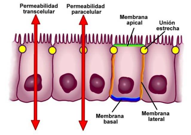
Tight junctions, also known in the literature as occlusive junctions, are sectors in the cell membranes of neighboring cells that are closely linked - as the name "tight junction" indicates.
Under average conditions, the cells are separated by a distance of 10-20 nm. However, in the case of tight junctions, this distance is significantly reduced and the membranes of both cells lead to touching or even merging..
A typical tight junction is located between the side walls of neighboring cells at a minimum distance from their apical surfaces..
In epithelial tissue, all cells make such junctions to stay together. In this interaction, cells are arranged in a pattern that resembles a ring. These unions cover the entire perimeter.
The tight contact regions are surrounding the entire cell surface. These regions form anastomosed contact strips of the transmembrane proteins known as occludin and claudin. The term anastomosis refers to the union of certain anatomical elements.
These two proteins belong to the group of tetraespanins. They are characterized by having four transmembrane domains, two outer loops, and two relatively short cytoplasmic tails..
Occludin has been shown to interact with four other protein molecules, called zonule occludin and abbreviated as ZO. This last group includes the proteins ZO 1, ZO 2, ZO 3 and afadin.
Claudin, for its part, is a family of 16 proteins that constitute a series of linear fibrils in tight junctions, which allows this junction to take on the role of a "barrier" in the paracellular pathway..
Nectins and junction adhesion molecules (JAM) also appear in tight junctions. These two molecules are found as homodimers in the intracellular space..
Nectins are connected to actin filaments through the protein afadin. The latter seems to be vital, since in the deletions of the gene that codes for afadin in rodents, they lead to the death of the embryo.
These types of junctions between cells perform two essential functions. The first is to determine the polarity of the cells in the epithelium, separating the apical domain from the basolateral domain and preventing undue diffusion of lipids, proteins, and other biomolecules from taking place..
As we mentioned in the definition, the cells of the epithelium are grouped in a ring. This structure separates the apical surface of the cell from the lateral and basal ones, which establishes the differentiation between the domains.
This separation is considered one of the most important concepts in the study of the physiology of epithelia..
Second, tight junctions prevent the free passage of substances through the epithelial cell layer, which translates into a barrier for the paracellular pathway..
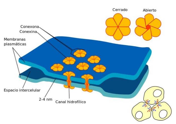
The gap junctions are found in regions devoid of limiting cytoplasmic membrane between neighboring cells. In a cleft junction, the cytoplasms of the cells connect and a physical connection is created where the passage of small molecules can occur..
This class of junctions is found in virtually all epithelia, and in other types of tissues, where they serve quite a variety of purposes..
For example, in various tissues cleft junctions can open or close in response to extracellular signals, as is the case with the neurotransmitter dopamine. The presence of this molecule reduces communication between a class of neurons in the retina, in response to increased light intensity..
The cleft junctions are made up of proteins called connexins. Thus, a "connexon" is obtained by the union of six connexin monomers. This structure is a hollow cylinder that is found crossing the cytoplasmic membrane.
The connexons are arranged in such a way that a conduit is created between the cytoplasms of adjacent cells. Also, the connexons tend to aggregate and form a kind of plates..
Thanks to the formation of these junctions, the movement of certain molecules between neighboring cells can occur. The size of the molecule to be transported is decisive, the optimal diameter is 1.2, as are calcium ions and cyclic adenosine monophosphate.
Specifically, they are inorganic ions and water-soluble molecules that can be transferred from one cell cytoplasm to the contiguous cytoplasm..
Calcium concentrations play a crucial role in this channel. When the calcium concentration increases, the axial ducts tend to close.
In this way, cleft junctions actively participate in the electrical and chemical coupling process between cells, as occurs in the muscle cells of the heart, which are responsible for transmitting electrical impulses..
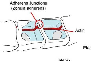
Below the tight joints, we find the anchor joints. Generally, these are located near the apical surface of the epithelium. In this group, we can distinguish three main groups, the zonula adherens or belt desmosome, the macula adherens or punctual desmosome and the desmosome.
In this type of junction, the adjacent cell membranes that are linked by zonules and adherent macules are separated by a relatively large cell distance - compared to the minimal space that exists in the case of tight junctions..
The intercellular space is occupied by proteins that belong to the family of cadherins, desmogleins and desmocholins attached to cytoplasmic plaques that present other proteins called desmoplakin, placoglobin and placophilin.
As in the case of tight joints, in the anchor joints we also observe the pattern of arrangement in the form of a ring or a belt. The zonula adherens is associated with actin microfilments, through the interaction of two proteins: cadherins and catenins..
In some cases, this structure is known simply as a desmosome, it is a punctiform union that is associated with intermediate filaments formed of keratin. In this context, these keratin structures are called "tonofilimanetos". The filaments extend from point to point in epithelial cells.
These provide strength and rigidity to the epithelial cells. Thus, it is believed that its main function is related to the strengthening and stabilization of adjacent cells..
Desmosomes can be compared to a kind of rivet or weld, as they resemble separate tiny dots and not continuous bands.
We find this type of junction in the intercalated discs that join the cardiocytes in the heart muscle and in the meninges that line the outer surface of the brain and spinal cord..
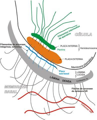
Hemidesmosomes fall into the category of asymmetric junctions. These structures have the function of anchoring the basal domain of the epithelial cell with the underlying basal lamina.
The term hemidesmosome is used because this structure appears, literally, "half" desmosome. However, from the point of view of their biochemical composition, both unions are totally different..
It is important to clarify that desmosomes are responsible for adhering one neighboring cell to another, while the function of the hemidesmosome is to unite the cell with the basal lamina.
Unlike the macula adherens or the desmosome, hemidesmosomes have a different structure, consisting of: a cytoplasmic lamina associated with intermediate filaments and a plate of external membranes, which is responsible for joining the hemidesmosome with the basal lamina, by means of a anchor filament.
One of the functions of hemidesmosomes is to increase the global stability of epithelial tissues, thanks to the presence of intermediate cytoskeletal filaments attached to the components of the basal lamina.
The plant kingdom lacks most of the cell junctions described above, with the exception of a functional counterpart reminiscent of cleft junctions..
In plants, the cytoplasms of adjacent cells are connected by pathways or channels called plasmodesmata..
This structure creates a continuum from one plant cell to the next. Although it differs structurally from cleft junctions, they have very similar roles, allowing the passage of small ions and molecules..
From a medical point of view, cell junctions are a relevant topic. Mutations in the genes that code for the proteins involved in the junctions have been found to translate into clinical pathologies.
For example, if there is a certain mutation in the gene that codes for a specific type of claudin (one of the proteins that mediates the interaction in tight junctions) it causes a rare disease in humans..
This is renal magnesium loss syndrome, and symptoms include low magnesium and seizures..
In addition, a mutation in the gene encoding the nectin 1 protein has been found to be responsible for cleft lip syndrome. This condition is considered one of the most common malformations in newborns..
Mutations in the nectin 1 gene have also been associated with another condition called ectodermal dysplasia that affects human skin, hair, nails, and teeth..
Pemphigus foliaceus is a blistering skin disease determined by autoantibodies against desmoglein 1, a key element that is responsible for maintaining the cohesiveness of the epidermis.


Yet No Comments