
The notion of extrapyramidal approach or the extrapyramidal system (EPS) arose as a result of anatomical and physiological studies aimed at understanding the way in which the central nervous system controlled the activity of the skeletal muscles, with the aim that the body assumed the appropriate body posture and produced the voluntary movements.
In this process, it was discovered that the control of muscle activity required control of the motor neurons of the anterior horn of the spinal cord, the only connection between the central nervous system and skeletal muscle fibers, and that this control was exercised by nerve projections from brain centers. superior.
Among these projections, an important pathway is formed by some axons that originate in the motor areas of the cerebral cortex and descend directly, that is, without scales, to the spinal cord, joining, as they pass through the medulla oblongata, in some prominences that due to their shape were given the name of "pyramids".
This tract was called the "pyramidal tract" or "corticospinal tract" and it was involved in the control of the fine and skillful movements executed by the distal portions of the limbs, while the existence of structures with motor function was recognized but not included. in this way (extra).
The term “extrapyramidal motor system”, already obsolete from the physiological point of view, is still used in clinical jargon to refer to those structures of the brain and brainstem that collaborate in motor control, but are not part of the pyramidal system or direct corticospinal.
Article index
The extrapyramidal pathway can be described as organized into two groups of components: one would be made up of a set of nuclei of the brain stem and its projections towards the spinal cord, and the other would be made up of the subcortical nuclei known as nuclei or basal ganglia..
In the brain stem there are groups of neurons whose axons project towards the gray matter of the spinal cord and which have been described as organized in two systems: one medial and the other lateral..
The medial system is made up of the vestibulospinal, reticulospinal and tectospinal tracts that descend through the ventral cords of the cord and exert control over the axial or trunk muscles, in addition to the proximal muscles of the extremities involved in body posture..
The most important component of the lateral system is the rubrospinal tract, whose axons project from the red midbrain nucleus, descend through the lateral cord of the cord and end up influencing the motor neurons that control the distal muscles of the extremities..
From the foregoing it can be deduced that the medial system collaborates in the basic postural adjustments, necessary for voluntary motor activity, while the lateral system deals, together with the direct corticospinal route, with the movements of the extremities aimed at a purpose such as reaching and manipulate objects.
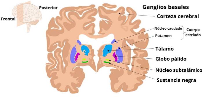
The basal ganglia are subcortical neuronal structures that are involved in the processing of motor information such as planning and programming of complex skillful movements, and whose alterations give clinical manifestations that are grouped into syndromes known as “extrapyramidal”.
Ganglia include the striatum, which is made up of the putamen and the caudate nucleus; the globe pallidus, which has an external portion (GPe) and an internal portion (GPi); the substantia nigra, organized into a compact portion (SNc) and a reticulated portion (SNr), and the subthalamic or Lewis nucleus.
These structures work by receiving information mainly from different regions of the cerebral cortex; information that activates internal circuits that affect an output neuronal activity that returns, via the motor portion of the thalamus, to the cerebral cortex.
Information about the ganglia enters through the striatum (caudate and putamen). From there, pathways that connect with the exit nuclei that are the GPi and the SNr start, whose axons go to the ventroanterior and ventrolateral nuclei of the thalamus, which, in turn, project to the cortex.
The different stages of the circuit are covered by neurons that belong to a particular neurochemical system and that can have an inhibitory or excitatory effect. The cortico-striated connections, the thalamic-cortical and the subthalamic fibers release glutamate and are excitatory.
Neurons whose axons exit the striatum use gamma amino butyric acid (GABA) as the main neurotransmitter and are inhibitory. There are two subpopulations: one synthesizes substance P as cotransmitter [GABA (+ Subst. P)] and the other enkephalin [GABA (+ Encef.)].
GABA (+ Sust. P) neurons have D1 dopamine receptors and are excited by dopamine (DA); they also establish a direct inhibitory connection with the outlets of the basal ganglia (GPi and SNr) that are also GABAergic but “+ dynorphin” and inhibit glutamatergic cells of thalamic-cortical projection.
GABA (+ Encef.) Neurons have dopamine D2 receptors and are inhibited by dopamine. They establish indirect excitatory connection with the outputs (GPi and SNr), since they project to the GPe inhibiting its GABAergic neurons, which inhibit the glutamatergic neurons of the subthalamic nucleus, whose function is to activate the outputs (GPi and SNr).
The compact part of the substantia nigra (SNc) has dopaminergic neurons (DA) that connect with the striatum making connections, as already mentioned, excitatory D1 on GABA cells (+ Sub. P) and inhibitory D2 on GABA cells (+ Encef .).
Then, and in accordance with the above, an activation of the direct pathway ends up inhibiting the outputs of the basal ganglia and releasing the activity in the thalamic-cortical connections, while the activation of the indirect pathway activates the outputs and reduces thalamic activity. -cortical.
Although the interactions and the exact joint functioning of the direct and indirect pathways just considered have not been clarified, the anatomical and neurochemical organization described helps us to understand, at least in part, some pathological conditions resulting from dysfunction of the basal ganglia..
Although the pathological processes that settle in the basal ganglia are diverse in nature and affect not only certain motor functions but also cognitive, associative and emotional functions, in clinical pictures motor alterations occupy a prominent place and most of the research has focused on them.
The movement disorders typical of basal ganglia dysfunction can be classified into one of three groups, namely:
- Hyperkinesias, such as Huntington's disease or chorea and hemibalism.
- Hypokinesias, such as Parkinson's disease.
- Dystonias, such as athetosis.
In general terms, it can be said that hyperkinetic disorders, characterized by excessive motor activity, present with a decrease in the inhibition that the outputs (GPi and SNr) exert on the thalamic-cortical projections, which become more active.
Hypokinetic disorders, on the other hand, are accompanied by an increase in this inhibition, with a reduction in thalamic-cortical activity.
It is a hyperkinetic disorder characterized by involuntary and spasmodic random shaking of the extremities and orofacial region, choreiform or "dance" movements that gradually increase and incapacitate the patient, speech disturbance and progressive development of dementia.
The disease is accompanied early by a degeneration of the GABA (+ Encef.) Striatal neurons of the indirect pathway.
As these neurons no longer inhibit GPe GABAergic neurons, they exaggerately inhibit the subthalamic nucleus, which stops exciting the inhibitory outputs (GPi and SNr) and the thalamic-cortical projections are disinhibited..
It consists of the violent contractions of the proximal muscles of the limbs, which are projected with force in movements of great amplitude. The damage in this case is the degeneration of the subthalamic nucleus, which results in something similar to that described for chorea, although not by hyper inhibition, but by destruction of the subthalamic nucleus.
It is characterized by difficulty and delay in the initiation of movements (akinesia), slowing of movements (hypokinesia), expressionless face or facial expression in a mask, gait alteration with decreased associated movements of the limbs during movement and tremor involuntary limb at rest.
The damage, in this case, consists of the degeneration of the nigrostriatal system, which are the dopaminergic projections that start from the compact region of the substantia nigra (CNS) and connect with the striatal neurons that give rise to the direct and indirect pathways..
The suppression of the excitation that the dopaminergic fibers exert on the GABA cells (+ Sust. P) of the direct pathway, removes the inhibition that these exert on the GABAergic outputs (GPi and SNr) towards the thalamus, which is now more inhibited. intensity. It is then a disinhibition of the outputs.
On the other hand, the suppression of the inhibitory activity that dopamine exerts on the GABA cells (+ Encef.) Of the indirect pathway releases them and increases the inhibition that they exert on the GABA cells of the GPe, which disinhibits the neurons of the nucleus subthalamic, which then hyperactivate the outputs.
As can be seen, the final result of the effects of dopaminergic degeneration on the two internal pathways, direct and indirect, is the same, whether it is disinhibition or stimulation of the GABAergic outputs (GPi and SNr) that inhibit the nuclei. thalamic and reduce their output into the cortex, which explains the hypokinesis

Yet No Comments