The lymphatic vessels they are transparent and gnarled ducts that have convergent branches. These lead to the veins the lymph and chyle (of intestinal origin). On their way, the lymphatic vessels traverse round adjoining structures known as lymph nodes..
The lymphatic vessels are also known as absorbent vessels and are found in all parts of the body, with the exception of the placenta and the central nervous system, which lack lymphatics..
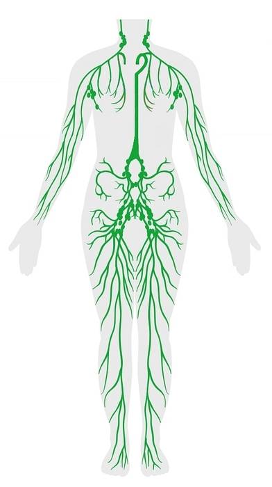
Like blood vessels, they have a tree-like or branched arrangement and are distributed in two planes: one superficial or subcutaneous (in the case of the extremities and the trunk) and the other deep or intramuscular..
The numerous valves that some of these vessels have, and the dilations on them, give them the appearance of a rosary. Lymphatic vessels on one side differ from those on the opposite side.
Article index
Lymphatic vessels originate in the tissues in tubules or capillaries with a blind end and a single cell layer.
These capillaries form a network that is drained by the lymphatic vessels, collecting trunks, and lymphatic ducts. They are more voluminous than blood capillaries and, as they extend proximally, their diameter gradually increases.
Its structure is similar to that of blood veins. It has walls with two membranes (external and internal) and a fibromuscular tunica media.
Lymphatic vessels have variable shapes and may have or lack valves. Valveless or "avalvular" lymphatic vessels are regular or rectilinear. Those vessels that present valves are irregular, showing alternately narrowing and dilatation, where the valves are implanted in pairs.
Valves are rare in the thoracic duct and in the descending vessels of the head and are formed mainly by invaginations of the internal tunica.
There may be cases of valve insufficiencies that lead to lymph reflux or stasis, which in turn generates edema of lymphatic origin. These vessels are contiguous with the veins and can be superficial or deep..
Lymphatic vessels thicken and decrease in number as they move away from their origin. During their journey they branch and rejoin each other or with adjacent branches, forming species of plexuses where they anastomose and distend.
After a more or less long journey, all the vessels branch out, appearing to end in the lymph nodes. Beyond these, they appear in the form of roots that meet similarly in the veins.
Some lymphatic vessels, such as those in the limbs, travel relatively long paths, without being interrupted by the nodes. In other vessels, such as those of the mesentery, ganglia are found in a continuous manner, fulfilling very short routes, while some pass close to ganglia without stopping in them.
After traveling more or less long trajectories, the vessels of the lower half of the body and of the upper and left quarter, end in an elongated trunk in the thoracic canal in the left subclavian vein. The vessels of the rest of the body terminate in a short trunk in the right subclavian vein.
The lymph is absorbed by the lymphatic networks and later by the lymphatic vessels. From these they enter the first ganglia, crossing the sinuses of said ganglia and undergoing a transformation. Subsequently it is directed towards the thoracic canal or the right thick lymphatic vessel, then spilling into the blood vessels at the base of the neck.
From the right supradiaphragmatic portion of the body, the lymph flows to the right lymphatic vessel, while the lymph from the left subdiaphragmatic and supradiaphragmatic portions, arrives through the thoracic canal in the left subclavial vein..
Superficial lymphatic vessels are found in subcutaneous tissues and skin, generally accompanying superficial veins. In certain places in the extremities the superficial vessels join the deep lymphatic vessels.
The superficial lymphatic vessels of the lower extremities drain by following the greater saphenous vein on the medial side, and the lesser saphenous vein on the lateral side. The drainage of the middle limbs coalesces with the superficial inguinal nodes around the great saphenous vein and around the saphenous hiatus.
Lymph from the lower part of the anal canal and the female genitalia are received by the horizontal group of inguinal nodes below the level of the umbilicus. The efferent vessels of the superficial inguinal ganglia pass through the cribriform fascia of the saphenous hiatus, ending in the external iliac ganglia.
Vessels connected to the lesser saphenous vein flow into the popliteal ganglia through the roof of the fascia.
The deep lymphatic vessels drain deep areas with respect to the fascia, accompanying the blood vessels of the region.
The deep lymphatic vessels follow the satellite veins, following the same path as the deep veins. These vessels are associated with small nodes. The anterior and posterior tibial vessels drain the lymph from the knees into the popliteal ganglia.
The vessels leading from the popliteal ganglia reach the deep inguinal nodes found on the medial side of the femoral vein. These ganglia also receive the deep vessels in the area of the femoral artery..
Lymphatic vessels exit the lower extremities from the deep and superficial inguinal nodes and reach the external iliac nodes.
The lymphatic vessels are responsible for transporting lymph, which is a clear liquid with high lipid content and also carries cells and debris or waste from the immune system.
Chyle, a milky-looking liquid compound, formed in the small intestine and made up of lipids, bile and pancreatic debris, is also transported by the lymphatic vessels. There are specific vessels that transport this material, and they are called chyliferous or lactiferous.
These two substances are transported to the trunks from their origins, and in the case of the lower extremities, the valves are in charge of maintaining this direction in the transport, avoiding the reflux or retrograde course of the liquids.
The main function of the lymphatic vessels is reduced to the absorption of liquids and substances dissolved in them, found in the interstitial spaces of the tissues and in the body cavities..
These vessels exert their action on ingested and fluidized food through digestion, liquid substances in contact with membranes, substances formed by the dissolution of organic tissues and blood plasma transudate through the walls of the vessels..
In the process of blood circulation, the absorption of plasma by the lymphatic vessels is vital. To maintain normal turgor in blood cases, lymphatic vessels must continuously absorb as much plasma as is produced by blood vessels..
If the lymphatic vessels do not absorb the plasma effectively, a state of dropsy occurs. This condition can also be generated by occlusion of the lymphatics, as in the case of phlegmasia alba disease and edema of the limbs due to obstruction caused by the absorption of an animal venom..
The lymph passes through the lymph nodes through the lymphatic vessels following a continuity between two types of vessels: afferent and efferent.
The afferent and efferent vessels lose their characteristics within the nodes, that is, they are not really lymphatic vessels as they enter the nodes. Between these types of vessels there are lymphatic sinuses, which are systems of lagoons that surround the follicles and lymphatic channels..
The lymphatic sinuses extend from the afferent to the efferent vessels, surrounding the follicles and lymphatic channels, separating the latter from the fibrous septa. These sinuses are traversed by the connective tissue filaments that extend from the follicles to the septa, forming a kind of covering on the follicles..
The lymph is received by the lymphatic sinuses and then transmitted to the efferent vessels.
The afferent lymphatic vessels are commonly numerous and branch in the peripheral zone of the node. When it is associated with the fibrous lining of the lymph node, its wall joins the connective tissue of said lining, opening at various outlets in the lymphatic sinuses that surround the follicles.
The afferent lymphatic vessels discharge the lymph on the surface of the follicles, circulating in the spaces between the follicles and the fibrous septa. Later it passes to the medullary layer, bathing the walls of the lymphatic channels and thus passing to the efferent channels.
Efferent vessels continue with lymphatic sinuses, making their origins difficult to recognize.
The lymph crosses the areolas of the sinuses of the medullary layer and reaches a conduit that is in the connective tissue of the stroma. Finally it empties from the depressed point, being able to differentiate one or more efferent vessels equipped with valves.
In the lymph nodes, there are no lymphatic vessels properly because, as mentioned, these vessels lose their characteristics.
Instead, thin epithelial cells have been observed in the walls of the lymphatic sinuses, septums, follicles, and node filaments. These cells appear to be in continuity with the internal cells of the lymphatic vessels..


Yet No Comments