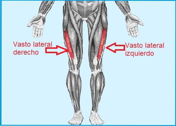
The vast lateral or vastus lateralis, as it is also known, is a superficial and palpable muscle, located in the anterolateral portion of the thigh. It is an even muscle, as there is one in each lower limb (leg). Its name comes from the Latin musculus vastus lateralis.
The vastus lateralis, together with the vastus medialis, vastus intermedia and rectus femoris make up the most robust muscle in the human body called the quadriceps, the vastus lateralis being the largest of the four.
This set of muscles act synergistically to make it possible to extend the knee joint. In addition, it also participates in the stability of the knee joint.
On the other hand, the hamstring muscles antagonize the action of the vastus lateralis and in general of the entire quadriceps, as they help in flexion of the knee joint, among other functions..
The vastus lateralis is a muscle that, because it does not have important blood vessels and sometimes does not have great innervation, is used for intramuscular self-injection. Although for this purpose it is preferred to choose the rectus femoris.
Article index
It is delimited on the medial part by the rectus femoris or rectus femoris muscle, while on the lateral part it is delimited by the iliotibial band, by the fascia lata and by the gluteus maximus..
The muscle can be palpated, for this the patient is asked to extend the leg straight and this will be noticed.
This muscle arises from the lower portion of the greater trochanter and the upper portion of the intertrochanteric line, and travels down the entire femur on its outer side.
Its fibers are arranged obliquely outwards and within 3/4 of their extension they are strongly attached to a fibrous collagen membrane called the aponeurosis, originating in the greater trochanter.
Below the muscle is another aponeurosis, from which many fibers of the vastus lateralis arise, as well as other nearby muscles, such as the gluteus maximus tendon and the lateral intermuscular septum..
During its course, its fibers have several points of insertion, specifically it is attached to the trifurcation and lateral lip of the linea aspera (superior 2/3), to the diaphysis of the femur in its superior anterolateral portion, in the fascia lata and, finally, in the lateral intermuscular septum.
Subsequently, the muscle passes over the lateral border of the patella and inserts into the tuberosity of the tibia, thanks to the patellar tendon. There it fuses with fibers from the rest of the muscles that make up the quadriceps. This provides a reinforcement of the capsule that lines the knee joint..
The vastus lateralis muscle receives a branch of the deep femoral artery called the lateral femoral circumflex artery.
The vastus lateralis muscle is innervated by the femoral nerve (L2-L4), like the rest of the quadriceps muscles, with the exception of the rectus femoris.
It is a great knee extensor. This is its main function, which fulfills in complete synergy with all the muscles that make up the muscle group called quadriceps. This means that the rest of the quadriceps muscles are vastus lateralis agonists..
In addition, it participates in the stability of the patella when it is flexed, since it generates a force posterior to it. All the muscles of the quadriceps and the patellar tendon participate in this action..
In this sense, the vastus lateralis exerts a lateral traction force on the patella, being counteracted by the vastus medialis. In this way the balance is achieved.
The extension of the knee allows us to position the lower limb in a straight way. This movement collaborates in actions such as getting up from the squatting position, standing, walking, running, jumping, among others..
The vastus muscle, like other muscles, is not exempt from stress and bruising. This could be injured if it is not properly warmed up before starting an exercise routine or as a result of an impact where the muscle is compressed against the bone.
The vastus lateralis is one of the muscles of the quadriceps that can cause a lot of pain, instability of the patella and even sleep disturbance, thanks to the presence of up to 5 key areas of trigger points.
Trigger points are distributed from the origin of the muscle to its insertion and are identified as PG1, PG2, PG3, PG4, and PG5. They can create referred pain towards the iliac crest or towards the knee, depending on the location of the pain point.
Trigger point 1 (PG1) affects the knee, producing a very pathognomonic symptom characterized by the sensation of having the patella stuck or blocked. That is, there is an impossibility of bending the knee and there is pain in the lateral border, which can extend upwards.
The PG2 produces more pain in the lateral area and this extends upwards. PG3 causes pain towards the posterolateral portion of the thigh and the popliteal fossa, that is, behind the knee (hamstrings).
In PG4 the pain refers more to the lateral part of the patella, with severe pain referred to the entire lateral aspect of the muscle. Finally, the PG5 is located towards the proximal portion of the muscle with localized pain referred to the iliac crest.
In PG4 and PG5 the pain is usually very intense, interrupting the patient's sleep, since it is impossible for the patient to lie on the affected side.
This clinical picture presents with repetitive dislocations at the level of the patella, which causes a lot of pain, discomfort, movement limitations and quadriceps muscle atrophy. Treatment for this condition is usually surgical..
With the patient completely straight lying on his back (supine position), the patient is instructed to contract the muscle within his possibilities, trying to hit the hamstring of the stretcher. The capacity of the contraction is evaluated.
The exercise called the sissy squat and its different variants are very useful to strengthen the muscles that make up the quadriceps.
This squat consists of opening the legs to the width of our hips, leaning on the tips of our feet. Then with the hands at the waist we pull ourselves back carefully, making the knees, hips and shoulders form a straight line. The knees are slightly bent and the back straight.
Variants of the sissy squat include the Roman chair and knee hinge..
There is a special machine to perform this exercise, although sometimes it can be improvised.
The idea is that you stand with your feet close together and planted on the ground. These will be adjusted by a kind of weight and at the level of the ankles there is a support that will hold you to prevent you from falling. In this position you should try to pull yourself back keeping your back always straight.
Basically the movement is the same as in the sissy squat exercise, but this time you will do it kneeling on a comfortable surface and not so low.


Yet No Comments