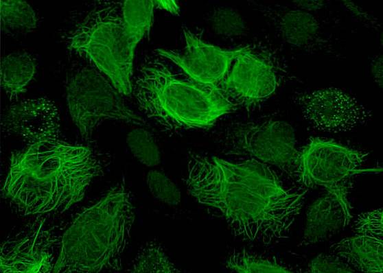
The vimentin it is one of the 57 kDa fibrous proteins that are part of the intracellular cytoskeleton. It is part of the so-called intermediate filaments and is the first of these elements to form in any type of eukaryotic cell. It is found mainly in embryonic cells, and remains in some adult cells, such as endothelial and blood cells..
For many years scientists believed that the cytosol was a kind of gel in which the cellular organelles floated and there were proteins in dilution. However, they now recognize that reality is more complex, and that proteins form a complex network of filaments and microtubules that they have called the cytoskeleton..
Article index
Vimentin is a fibrous intermediate filament protein, 57kDa and contains 466 amino acids. It is common as part of the cytoskeleton of mesenchymal, embryonic, endothelial, and vascular cells. It is rare to find this protein in non-eukaryotic organisms, but it has nevertheless been isolated in some bacteria.
Vimentin is laterally or terminally attached to the endoplasmic reticulum, mitochondria, and nucleus.
In vertebrate organisms, vimentin is a highly conserved protein and is closely related to the immune response and the control and transport of low-density lipids.
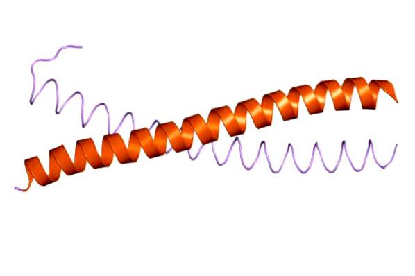
Vimentin is a simple molecule that, like all intermediate filaments, has a central alpha-helical domain. At its ends (tail and head) it has amino (head) and carboxyl (tail) domains without helixes or non-helical ones..
Alpha-helical sequences present a pattern of hydrophobic amino acids, which serve or contribute to the formation of the hydrophobic seal on the helical surface..
As its name implies, it is the structural support of eukaryotic cells. It goes from the inner face of the plasma membrane to the nucleus. In addition to serving as a skeleton, allowing cells to acquire and maintain their shape, it has other important functions.
Among these is participating in cell movement, as well as in its division process. It also supports intracellular organelles and allows them to actively move within the cytosol, and participates in some intercellular junctions..
In addition, some researchers argue that the enzymes believed to be in solution in the cytosol are actually anchored to the cytoskeleton, and enzymes of the same metabolic pathway must be located close to each other.
The cytoskeleton has three main structural elements: microtubules, microfilaments, and intermediate filaments. These elements are found only in eukaryotic cells. Each of these elements has a characteristic size, structure and intracellular distribution, and each also has a different composition.
Microtubules are composed of tubulin heterodimers. They have a tubular shape, hence their name, with a diameter of 25 nm and a hollow center. They are the largest elements of the cytoskeleton. Its length varies between less than 200 nm and several micrometers long.
Its wall is generally made up of 13 protofilaments, arranged around the central lumen (hollow). There are two groups of microtubules: on the one hand, the microtubules of the axoneme, related to the movement of cilia and flagella. On the other hand, are the cytoplasmic microtubules.
The latter have various functions, including organizing and maintaining the shape of animal cells, as well as the axons of nerve cells. They are also involved in the formation of mitotic and meiotic spindles during cell divisions, and in the orientation and movement of vesicles and other organelles..
They are filaments made up of actin, a protein of 375 amino acids and a molecular weight of about 42 kDa. These filaments have a diameter less than a third of the diameter of microtubules (7 nm), which makes them the smallest filaments of the cytoskeleton..
They are present in most eukaryotic cells and have various functions; among them, participate in the development and maintenance of the cellular form. In addition, they participate in locomotor activities, both amoeboid movement, and in muscle contractions, by interaction with myosin.
During cytokinesis (cytoplasmic division), they are responsible for producing segmentation grooves. Finally, they also participate in cell-cell and cell-extracellular matrix junctions..

With an approximate diameter of 12 nm, the intermediate filaments are the ones with the greatest stability and are also the least soluble of the elements that make up the cytoskeleton. Only found in multicellular organisms.
Its name is due to the fact that its size is between that of microtubules and microfilaments, as well as between those of actin and myosin filaments in muscles. They can be found individually or in groups forming bundles.
They are made up of a main protein, and various accessory proteins. These proteins are specific to each tissue. Intermediate filaments are only found in multicellular organisms, and unlike microtubules and microfilaments, they have a very different amino acid sequence from one tissue to another..
Based on the type of cell and / or tissue where they are found, the intermediate filaments are grouped into six classes.
Formed by acid cytokeratins that give mechanical resistance to epithelial tissue. Its molecular weight is 40-56.5 kDa
It is made up of the basic cytokeratins, which are slightly heavier than the previous ones (53-67 kDa), and help them to give mechanical resistance to the epithelial tissue..
Represented by vimentin, desmin, and GFA protein, which are mainly found in mesenchymal cells (as mentioned before), embryonic and muscle cells, respectively. They help give each of these cells their characteristic shape.
They are the proteins of neurofilaments. In addition to stiffening the axons of nerve cells, they also determine the size of these.
Represented by the laminae that form the nuclear scaffold (nuclear laminae). They are present in all types of cells
Formed by nestin, a 240 kDa molecule found in nerve stem cells and whose function remains unknown.
Vimentin participates in many physiological processes, but it mainly stands out for allowing rigidity and resistance to the cells that contain it, avoiding cell damage. They retain organelles in the cytosol. They are also involved in cell attachment, migration, and signaling..
Medical studies indicate that vimentin acts as a marker of cells derived from mesenchyme, during the normal and progressive development of cancer metastasis.
Other studies indicate that antibodies or immune cells that contain the VIM gene (the gene that codes for vimentin), can be used as markers in histopathology and often to detect epithelial and mesenchymal tumors..
The pharmaceutical and biotechnology industries have widely taken advantage of the properties of vimentin and used it for the production of an important variety of products such as genetically engineered antibodies, vimentin proteins, ELISA kits, and complementary DNA products, among many others..

Yet No Comments