
The vinagrillos, Also known as uropygians, they are a group of arachnid animals that belong to the order Uropygi and that are characterized mainly by having a flagellum at the terminal end of their prosoma, as well as anal glands that secrete a liquid similar to vinegar..
They were first described by the English zoologist Octavius Pickard Cambridge in 1872. They look fearsome, but are generally totally harmless. It is believed, according to the collected fossil records, that they originated in the Paleozoic era, specifically in the Carboniferous period and that they include more than 280 species.
Article index
The uropygians, as it happens with all the members of the animalia kingdom, are multicellular eukaryotic organisms.
In addition to this, they are triblastic and protostome. This implies that during their embryonic development they present three germ layers: ectoderm, mesoderm and endoderm. From them, each and every one of the specialized structures that will make up the adult individual are generated..
An important element is that, from an embryonic structure (blastopore), the mouth and anus of the animal originate simultaneously.
Similarly, uropygians are dioecious animals. This means that the sexes are separate. That is, there are female individuals and male individuals.
These arachnids also present bilateral symmetry, evidenced by drawing an imaginary line along the longitudinal plane of the animal and thus obtaining two exactly equal halves..
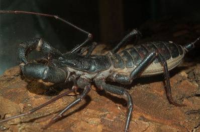
One of the most distinctive characteristic elements of the uropygians is that the males have glands at the level of the terminal segment of the prosoma that flow on both sides of the anus. These glands synthesize a substance that contains a high content of acetic acid and, therefore, smells like vinegar..
This liquid is used by these animals to defend themselves from possible predators or also to facilitate the process of capturing their prey. For human beings it is totally harmless.ç
The taxonomic classification of the vinagrillo or vinagrón is the following:
Domain: Eukarya
Animalia Kingdom
Phylum: Arthropoda
Subphylum: Chelicerata
Class: Arachnida
Superorder: Tetrapulmonary
Order: Uropygi.
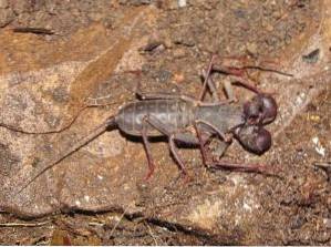
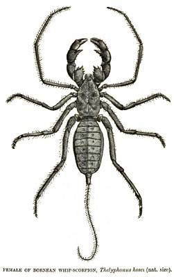
As with the rest of arachnids, uropygians have a body divided into two segments or tagmas: the cephalothorax (also known as the prosoma) and the abdomen (opistosome). They can measure up to 15 cm in length.
The characteristic element of uropygians, as far as morphology is concerned, is the flagellum that is found at the rear end of their body. The body is flattened dorsally and is typically dark reddish brown in color. They are small in size, although species that reach almost 8 cm have been described.
It is the anterior segment of the animal. It is covered by a kind of resistant shell or cuticle that serves as a protective shield for the uropygium..
The organs of sight are located on the dorsal surface of the prosoma, represented by a pair of simple eyes. In addition, there are three ocelli that have a lateral location. The ventral portion of the prosoma is entirely occupied by the first joint (coxa) of the legs..
In turn, the prosoma is where the animal's articulated appendages originate: two chelicerae, two pedipalps and eight legs..
They constitute the first pair of jointed appendages of the animal. They are made up of two pieces and are small in size. The proximal joint is stem-shaped, while the distal joint is nail-shaped..
They are widely developed. They have a clamp-shaped termination. They also present a series of very noticeable protrusions, which serve to capture the prey and be able to crush them..
The tweezers are made up of a mobile finger and a fixed finger. The first is made up of the tarsus and the basitarsus, while the fixed finger constitutes a projection of the arch called the tibia..
It is important to note that another protuberance is seen in the joint that corresponds to the patella, which, in general, constitutes another clamp..
In this sense, the pedipalps of the uropygians are one of the most prominent and developed of all the arachnids..
The locomotor appendages of the uropygians are eight and are distributed in pairs. They are thin in build and fragile in appearance, especially the first pair. More than a locomotive function, this first pair has a sensory function, since it is in charge of supplying the animal with information about the environment in which it is found..
The three remaining pairs of appendages fulfill the function of locomotion and movement of the animal. They also have, although to a lesser extent, some sensory structures such as trichobotrians..
It is the longest part of the animal. It is attached to the prosome by a structure called the pedicel. Similarly, according to some specialists, the opistosoma is divided into two areas or zones: the mesosome and the metasoma..
The mesosome is anteriorly located and encompasses nine of the twelve segments of the opistosome. It is in this sector where the holes corresponding to the reproductive system are located (in the second segment), as well as the spiracles that belong to the respiratory system (lateral position)..
The metasoma encompasses the last three segments of the opistosome. In its terminal segment is the anal orifice. On both sides of this, the orifices of the so-called anal glands are located.
Likewise, at the lateral and dorsal level of this last segment it is possible to observe small pale colored spots (omatoid). The function of these has not been demonstrated. However, they are used to differentiate one species from another..
A long, thin flagellar structure that is multi-articulated emerges from the posterior end of the metasoma. The function of this structure has to do with the release of the substance secreted by the anal glands for their protection. In addition, it constitutes a distinctive characteristic element of uropygians.
The uropygians have a complete digestive system, like the rest of the arachnids. This is made up of an initial area, known as the stomodeus, which corresponds to the orifice, the oral cavity and the esophagus..
This is followed by the midgut, also known as the midgut, and finally the proctodeum that culminates in the anal orifice..
The digestive system of this animal also has an attached organ, the hepatopancreas, which has to do with the storage of nutrients.
It is similar to other arachnids. It is made up of the so-called Malpighi tubes and also nephrocytes, which are responsible for collecting all the waste. The latter specialize in the storage of waste substances, while the Malpighi tubes lead to the proctodeo.
On the other hand, the coxal glands are also part of the excretory system. They owe their name to the fact that they flow right at the level of the first joint (coxa) of the last pair of the animal's legs..
It is made up of nerve clusters that together make up the ganglia. These are distributed throughout the body. Mainly associated with the organs of the digestive system such as the esophagus.
They present a ganglion at the level of the prosome, which fulfills, to a certain extent, the functions of a primitive brain. This emits nerve fibers to the simple eyes of the animal, as well as to the rest of the ganglia in the body.
Uropygians have a respiratory system made up of two types of structures: tracheas and book lungs..
The tracheae are defined as a set of tubes that branch into the interior of the animal into smaller ones called trachealas. These do not reach the cells of the animal directly as occurs in other arthropods, but instead lead to organs specialized in gas exchange: the book lungs..
These are made up of a series of lamellae, stacked one on top of the other, which resemble the pages of a book. Hence its name. In them the gas exchange is carried out.
The tracheas communicate with the exterior, through the spiracles that open towards the lateral portion of the opistosoma.
Uropygians are found primarily in moisture-rich ecosystems, such as those located in the tropics or subtropics. They are animals that prefer humid and dark places, which is why they are normally found under rocks, in caves and even buried in the ground..
These animals are purely carnivorous. They feed on small prey such as insects, amphibians and even other arachnids, including scorpions and spiders. In the capture process they use pedipalps which, due to their robustness, are ideal for this..
The type of digestion that uropygians have is external. This means that, by not being able to fully ingest the prey, they secrete a substance made up of digestive enzymes that pre-digestion the food, turning it into a kind of porridge..
The animal ingests this porridge and it is further degraded thanks to the action of digestive enzymes. Subsequently, in the mesodeum the necessary nutrients are absorbed and finally the waste products are released by the anus.
The reproduction of uropygians is characterized by being sexual, having internal fertilization, being oviparous and involving direct development.
In this sense, it is well known that sexual reproduction involves the fusion of male and female sexual gametes. Likewise, for the union of these gametes to occur, it is not necessary for a copulation process to occur..
The male releases a structure known as the spermatophore, in which the sperm are contained. Then, the female picks it up and introduces it, thus fertilization occurs. Later, the female lays the eggs in a site excavated by her in the ground.
Once the necessary time elapses, the young hatch from the eggs, which are attached to the mother's abdomen until they experience the first molt. Eventually they detach and subsist on their own. Throughout their life they will experience three more molts, after which they reach maturity.


Yet No Comments