
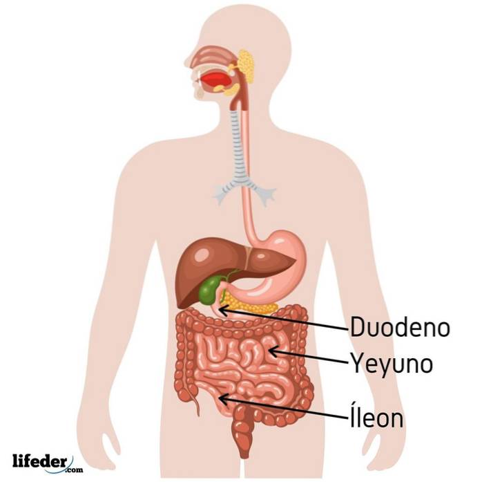
The jejunum is one of the three segments into which the small intestine of mammals and most vertebrate animals is divided, the other two being the duodenum and ileum..
The small intestine is part of the animal digestive system and, accompanied by the large intestine, fulfills the primary function of digestion, absorption and excretion of food ingested and initially processed in the mouth (with teeth, tongue and saliva) and then in the stomach (with gastric acids).
The small intestine is the longest organ in the gastrointestinal tract of animals, being approximately 7 meters long. It is divided into three sections:
Throughout this muscular tube, nutrients derived from food are digested and absorbed, the balance of water and electrolytes is maintained, an immune barrier is established against potential pathogens that enter the digestive tract and some hormones are produced..
All this is achieved, in part, thanks to a series of internal folds or structures (microvilli) that increase the total surface area of this organ many times, and also thanks to its particular anatomical and physiological characteristics.
While enzymatic digestion of food occurs in the duodenum, in the jejunum and ileum the components of these foods, vitamins, minerals and part of the water are absorbed (which is absorbed especially in the large intestine).
The jejunum is the middle portion of the small intestine. It is considered an intraperitoneal intestinal portion, which means that it is located within the membrane that covers the organs contained in the abdominal cavity (the peritoneum).
It represents approximately two fifths of the total length of the small intestine, being a region shorter than the ileum, but much longer than the duodenum.
Peristaltic intestinal movements are more vigorous and rapid during the transit of digested food along the jejunum, as this is where most of the intestinal absorption of nutrients occurs.
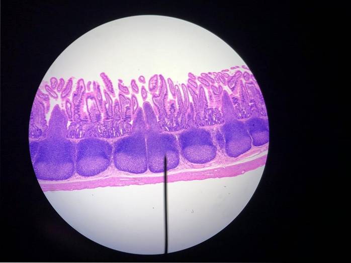
This portion of the small intestine has a neutral or slightly alkaline pH, between 7 and 8. It is covered by a layer of cells known as enterocytes, which are the ones that line the microvilli where the last stages of digestion take place and part of intestinal absorption.
The jejunum receives the nutritional material that has been previously processed in the mouth, stomach and duodenum.
Food intake begins with the mouth, where it is mechanically processed with the teeth and mixed with saliva, which contains some enzymes that initiate the digestion of some of the macromolecules contained in food, forming the food bolus..
The food bolus reaches the stomach, the hollow muscular organ located in the anterior portion of the abdominal cavity, where the chemical digestion of food begins, as large amounts of gastric juices rich in hydrochloric acid and other acidic substances and enzymes that favor the decomposition of food, forming a paste known as chyme.
Thanks to the muscular movements of the stomach, the chyme travels to the duodenum, where enzymes and other substances necessary for the digestion of proteins, carbohydrates and fats are released from the liver and pancreas..
The product of duodenal processing travels through peristaltic movements to the jejunum, where a fundamental process known as the absorption of nutrients occurs, specifically of sugars, fatty acids and amino acids derived from carbohydrates, fats and proteins digested in the duodenum..
Nutrients absorbed in the jejunum rapidly enter the bloodstream, from where they are distributed throughout all cells, tissues, and organs of the animal body, as the digested material continues its journey to the ileum and subsequently to the large intestine..
The jejunum is located, specifically, between the portions corresponding to the duodenum and the ileum, and corresponds to more than two-fifths of this organ, since it measures just under 2.5 meters long.
It begins in one of the distal regions of the duodenum known as the duodenojejunal flexure, which is actually anatomically indistinguishable from the jejunum.
It is located in the central part of the abdomen and many authors establish that there is also no distinguishable anatomical mark between the end of the jejunum and the beginning of the ileum..
The entire small intestine is made up of a wall made up of four layers: the mucosa, the submucosa, the muscularis propria, and the serosa..
Of these, the mucosa is the innermost layer, the one in direct contact with the food that enters the intestine through the stomach pylorus. The mucous layer is made up of three layers: the epithelium, the lamina propria, and the muscularis mucosa; and is the place where absorption occurs at the level of the jejunum.
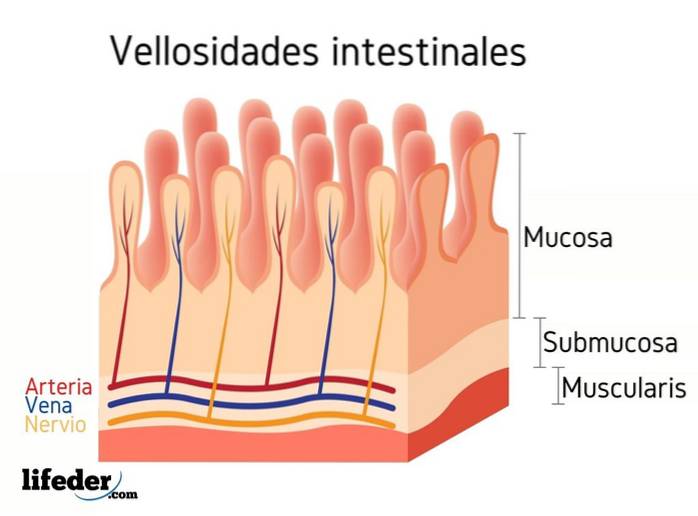
The submucosal layer is a dense layer, made up of connective tissue. It is perhaps the strongest layer of the intestinal wall and is the site where different glands, nerves and the blood and lymphatic vessels that supply the walls of the intestine meet..
The muscularis propria layer is made up of two layers of smooth muscle, an outer layer arranged longitudinally and an inner layer arranged in a circular manner..
The serous layer is the most superficial layer of the small intestine and is made up of a single line of mesothelial cells..
Compared to the duodenum and ileum, the jejunum has a thicker mucosal lining, a thicker muscle wall, a larger diameter, and less mesenteric fat; in addition, the veins that irrigate it are longer.
The jejunum, as well as the rest of the small intestine, has an inner surface that is lined with a series of transverse folds formed by the mucosa and submucosa layer, which increase the surface area and facilitate the intestinal absorption of nutrients..


Yet No Comments