
The microscopic anatomy It is the science that studies the tiny structure of cells and tissues that make up the body of organisms. To be able to develop effectively, this discipline requires precision tools, such as the ultraviolet light microscope or the electron microscope..
Therefore, it can be said that this science made great progress during the second half of the 19th century, since in this period optical microscopes were perfected. This allowed the development of new methods that facilitated the study of tissues..
Starting in the 20th century, microscopic anatomy expanded its knowledge thanks to the development of microscopy tools, which obtained greater power of magnification and resolution, achieved through technological advances. In addition, laboratory techniques were also refined, which facilitated observation.
It is important to highlight that two important scientific branches derive from this discipline, such as histology and cytology. The first studies the composition of organic tissues, focusing on the interior of cells and corpuscles; the second is also dedicated to the study of cells, but from a structural, biochemical and physiological level.
Article index
The history of microscopic anatomy begins with the appearance of general anatomy, whose origins were in turn linked to the beginnings of medicine. According to the author Clara García Barrios, in her text Origin and history of anatomical dissection (1999), the first anatomical remains began with the search to preserve human corpses.
Consequently, through embalming, mummification, and other preservation techniques, humans began to become familiar with body tissues. These techniques come from very remote civilizations, such as the ancient Egyptians or the Inca civilization.
It should be noted that to mummify and embalm it was necessary to make cuts, separate structures and access cavities, giving rise to the concept of dissection, which founded the bases of all anatomical sciences..
Anatomy as a science was born with the ancient Greeks. One of the most eminent physicians of this period was Hippocrates (460-370 BC), who is considered the father of medicine. Later, Aristotle (384-322 BC) was able to distinguish the nerves, tendons, bones and cartilage in the body of animals.
In the Alexandrian period, Herófilo (335-280 BC) practiced the first dissection of human corpses, giving rise to the concept of anatomy, which means "I short", in ancient Greek. This doctor discovered several anatomical formations, such as the brain and its meninges, the nerves, the milk vessels, the prostate and the duodenum..
Later, Erasistratus (350-300) considered the possibility that the organism was made up of tiny and invisible particles. This thought gave rise to what would later become microscopic anatomy..
The first scientist to observe cells was Robert Hooke in 1665, who managed to describe and draw the dead cells present in a cork; this he achieved by using a very primitive microscope. However, it was Antony Van Leeuwenhoek (1632-1723) who first observed a group of living cells.
In order to carry out his observations, Leeuwenhoek built a series of rather rudimentary but very successful microscopes for the moment, which allowed him to describe the cells present in blood and algae. His work was only descriptive, however, it served to discover the complex microscopic world.
The word "anatomy" comes from the Greek "anatomy", Which can be translated as" dissection ", although it also means" I cut ". Therefore, it can be established that anatomy is a science in charge of studying the shapes and structures of body parts, both of humans and animals..
As for the word “microscopic”, it comes from the noun “microscope”, formed by the Greek roots “micro” and “scopio”, which respectively mean “small” and “look”. Therefore, this word refers to the action of observing something that is very small.
In conclusion, the goal of microscopic anatomy is to examine biological structures that cannot be seen without being magnified. Through magnifying glasses, the scientist can reveal aspects that escape the human eye; the more advanced the microscope, the greater the amount of detail that cells and tissues present.
In order to carry out its investigations, microscopic anatomy requires the techniques of the microscope. One of the microscopes most used by scientists is the fluorescence light microscope, which uses quartz crystals and produces illumination through mercury lamps. This tool does not use filters and the results must be observed on photographic plates.

This instrument is essential when studying microscopic anatomy. It works in a similar way to a spectrophotometer, however, it differs from this because the results are recorded in photographic images.
The final result cannot be observed directly by the eyepiece since ultraviolet light can damage the researcher's retina. This method facilitates the detection of acids and proteins; also allows the obtaining of RNA from cells.
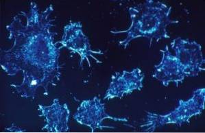
Electron microscopes are the most used today by this discipline. It differs from the previous ones in the fact that it uses electrons instead of using visible light to get images of tiny elements..
The first electron specimen was designed by Max Knoll and Ernst Ruska in 1925 and currently there are two types: transmission electron microscopes and scanning electron microscopes..
Microscopic anatomy uses other scientific branches to be able to develop its investigations more efficiently, these being histology and cytology. Although both disciplines are focused on different objectives, both agree that they require the use of a microscope to be carried out..
Histology allows microscopic anatomy to know the alveolar membranes present in various tissues of the body, while cytology provides in-depth knowledge about cells, both in their normal state and in a possible pathological state..


Yet No Comments