The appendectomy It consists of a surgical intervention aimed at the removal of the inflamed cecal appendix. This procedure is performed as an emergency for appendicitis, taking into account the risk that this disease represents. It is the most common surgery today.
The knowledge and development of appendectomy as a surgical technique occurred between the 18th and 19th centuries. The first appendix surgery on record occurred in 1735, performed by Amyan, a military surgeon. It is between the middle and the end of the 19th century when the technique and diagnostic procedures of appendicitis are documented..
The appendix is a structure located in the cecum, a portion of the large intestine. Organ function has been related to immune activity, but it is not a vital structure. Appendicitis is the inflammation of the appendix mainly due to obstruction mechanisms. This condition, although common, is potentially serious.
Appendicitis presents symptoms that guide its diagnosis, such as abdominal pain, loss of appetite, nausea, vomiting and, occasionally, fever. The pain classically begins in the upper hemiabdomen, and then radiates and locates in the right iliac fossa. Depending on the time of evolution, the appendix can be perforated and produce peritonitis.
In addition to the clinical examination, the diagnostic approach to appendicitis includes the performance of laboratory tests, radiology and ultrasound. An above-normal white blood cell count or imaging evidence will confirm the diagnosis of appendicitis..
Once the diagnosis of appendicitis is made, the treatment of choice is appendectomy. On occasion, preventive removal of the healthy appendix can occur during a laparotomy. The prophylactic appendectomy is performed, prior knowledge of the patient, to avoid future surgeries.
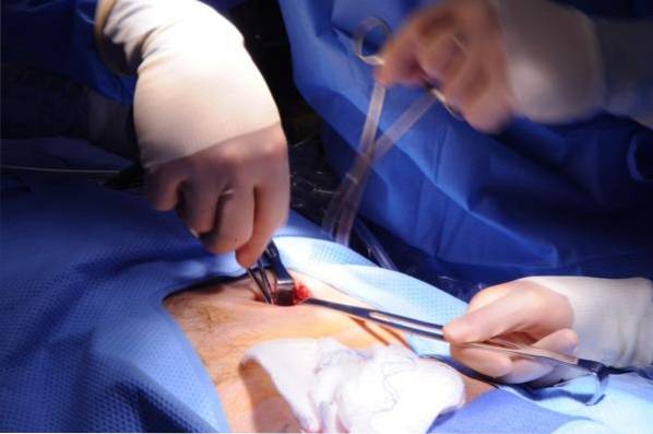
Open or traditional appendectomy is the most used, and consists of a surgical approach through an incision in the abdominal wall. Laparoscopic surgery is an instrumental technique that represents an option to perform appendectomy..
Article index
The only justification for performing an appendectomy is the unequivocal diagnosis of appendicitis. Considering the implications that surgery has for a patient, there must be an adequate prior diagnostic basis. History, precise clinical examination, and laboratory and imaging examinations are essential tools.
There are two procedures to perform an appendectomy: a traditional technique, or open appendectomy; and the laparoscopic approach.
The traditional and most common procedure used is open appendectomy. It can be performed with the patient under general or epidural anesthesia, according to the complexity of the surgery. This technique consists of several phases:
It consists of the adaptation of the patient to the surgical act. The operative area is the lower right quadrant of the abdomen, topographic location of the appendix.
First, with the operative area shaved, we proceed to rigorous cleaning with antiseptics. Once clean, the area is delimited with the placement of sterile material, fields and sheets.
The umlaut is the separation of tissues through incisions and cuts. To locate the appendix in the abdomen, the umlaut should be performed in different planes from the outside to the inside: skin, muscular aponeurosis, muscle and peritoneum. This procedure is done with the use of scalpel, forceps, scissors and special spacers..
- The initial incision will depend on the clinical phase of appendicitis, time of evolution and decision of the surgeon. The most commonly used incisions are McBourney's oblique, Lanz's paramedial oblique, and the right infraumbilical pararectalis. The pararectalis is usually used when there is suspicion of complications, being the easiest to extend, if required.
- The most widely used McBourney technique provides an overview of the surgical procedure. An oblique incision is made in the skin, just in the outer third of a line drawn from the umbilicus to the right iliac crest. To perform it, a conventional scalpel and an electrocautery are used, for cutting and cauterization.
- Once the skin is separated, the muscular aponeurosis is exposed, which will be cut and separated with the use of scissors and forceps. The oblique muscle is separated according to the direction of the fibers, without cutting. When separating the muscular plane, the transverse fascia and the peritoneum are observed, the cut of which will expose the abdominal cavity.
- The first inspection of the abdominal cavity will show if there is any abnormal fluid, either pus or blood. The portion of the colon, the cecum, is located to find the cecal appendix manually or instrumentally. When the appendix is exposed, its appearance -including its attachment to the colon- and that of neighboring structures are reviewed.
- The position of the appendix with respect to the cecum is inferior and slightly posterior. The positional variants can be lateral, pelvic and retrocecal, assuming different degrees of complexity in the technique. The search for a posterior or retrocecal appendix is more laborious.
- Removal of the appendix consists of several phases. The first phase consists of locating the appendicular artery located in its support structure (the mesoappendix) and ligating it. The second phase involves the double ligation, proximal and distal, of the appendicular base. Finally, the cut will be made with a scalpel soaked in iodine between the two ligatures..
- When the tissue of the stump and the appendicular base is very damaged, the surgeon opts for invagination of these. Invaginate the stump consists of introducing this structure into the healthy tissue of the cecum, and closing it by means of non-absorbable sutures. It is a technique used in cases of perforated or gangrenous appendicitis.
A thorough review of the abdominal cavity is necessary before finalizing the intervention. Verification of ligatures, active bleeding, existence of surgical medical material and examination of organs are part of this review. The operation culminates with washing and aspiration of the abdominal cavity using saline solution..
The closure of the operative area constitutes the structural restitution of the separated planes in the umlaut. The synthesis of the tissues will be carried out using suture thread or staples, appropriate to the tissue.
The suture will be from the deepest to the superficial plane: peritoneum, aponeurosis, muscle, muscle fascia, subcutaneous cellular tissue and skin.
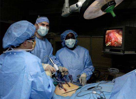
Laparoscopy is a minimally invasive technique, based on the use of a video camera and special instruments for the abdominal surgical approach. The use of laparoscopy in appendectomy depends both on the availability of specialized equipment and personnel and the absence of contraindications.
The indication for laparoscopic appendectomy is related to the patient's medical conditions. Hemodynamic instability, peritonitis, abdominal distension, extreme obesity, respiratory disease, pregnancy and previous abdominal surgeries are contraindications for its use..
The phases of laparoscopic surgery are similar to open surgery. Patient preparation, diaeresis by planes, extraction of the appendix, revision and closure by planes are carried out, although with obvious differences.
- Cleaning of the operative area with antiseptics and placement of sterile drapes.
- The type of anesthesia of choice in general is inhalational.
- The umlaut by planes is intended to allow the introduction of trocars or portals for the video camera and instruments. Usually two or three 2 cm incisions are made in the abdominal wall.
- The abdominal cavity must be insufflated with carbon dioxide to enlarge it and allow the visualization of the structures and mobility of the instruments..
- The instruments used, such as cautery, forceps and scissors, are adapted for the technique. The ligatures of the appendix and its meso are made through the use of ligatures and special staples..
- The final check-up is done by chamber examination, flushing, and saline aspiration. The removal of the trocars precedes the closure by planes of the incisions.
Open appendectomy continues to be the most widely used today; however, laparoscopy is an acceptable alternative.
Although more expensive than traditional surgery, the cost-benefit ratio is higher than this. Recovery of the laparoscopic patient is faster.
The success of appendectomy depends on both the results of the surgery and the patient's recovery. Factors such as the general condition of the individual, the surgery performed and the reaction to the procedure influence recovery.
Postoperative care serves to prevent complications and decrease hospitalization time. In uncomplicated appendectomies, in-hospital surveillance will be 24 to 48 hours.
After surgery, the effect of anesthetics must be completely reversed. In the immediate postoperative period, the prevention of possible reactions to anesthesia is carried out in the recovery room. It is the responsibility of the anesthesiologist to control and monitor the complete recovery of the patient..
Monitoring vital signs - such as heart rate, blood pressure, and respiration - can alert you to early complications.
Body temperature is measured regularly to detect the presence of fever. Stability of vital signs is a criterion for the absence of complications and recovery after surgery.
All abdominal surgery involves a period of rest from intestinal activity. The patient must maintain an absolute diet until the recovery of normal movements of the digestive system. Once indicated, a liquid diet will be started, followed by soft foods.
After an appendectomy, abundant foods, legumes or foods that promote abdominal distension should be avoided.
Abdominal surgeries carry a potential risk of intra-abdominal or operative wound infection. The use of antibiotics is a measure to avoid infections in the postoperative period of appendectomy.
Pain after appendectomy is common. Postoperative catheter analgesia systems are an option in cases of severe pain.
During the hospitalization period, intravenous analgesics are used to treat episodes of pain secondary to surgical instrumentation. Oral pain relievers are for outpatient use.
One of the measures to prevent infections in the operative wound is cleaning it, which must be done daily. The first days the operative area should be covered with sterile dressings..
Regular medical consultations are a surveillance measure during the mediate postoperative period. Outpatient medical check-ups are intended to assess the patient's health and proper wound healing. Late complications can be detected during regular check-ups.
Complications of an appendectomy can stem from the surgery, the stage of the appendicitis, the physical conditions of the patient, or failures in postoperative care. These complications can appear early or be late consequences.
The most common complications are those caused by wound or intra-abdominal infections. Other complications that occur can be intra-abdominal hemorrhages, accidental organ damage, and leakage of intestinal contents due to loss of ligatures of the appendicular stump or necrosis of the cecum..
Infections are due to bacterial contamination of the abdominal cavity and wound. The presence of germs, especially bacteria, can occur due to the use of non-sterile material, intraoperative contamination, or leakage of intestinal bacteria in cases of perforated or gangrenous appendicitis.
Among the most frequent infections are intra-abdominal abscesses and abdominal wall abscesses..
An infectious complication involves the stay of the patient in the hospital. The use of antibiotics, drainage of the abscess and cleaning of the operative wound are the measures to treat this complication..
Intra-abdominal bleeds occur due to bleeding vessels due to careless hemostasis or loss of blood vessel ligation. Accidental organ injury can cause bleeding.
Free blood in the abdominal cavity irritates the peritoneum producing severe pain and, depending on the volume of blood lost, signs of hypovolemic shock. Bleeding in the abdominal cavity requires surgery to locate the source of the bleeding and repair it.
In the development of an appendectomy, accidental injury to organs adjacent to the appendix is possible. An organ injury should be treated as soon as it is detected and, if it is significant, it will warrant surgery.
- Foreign bodies, consisting of medical material, accidentally left in the abdominal cavity will produce inflammatory reactions, serious infections and pain.
- The use of a bladder catheter during the operation can cause urethral injuries or urinary tract infections, being a minor complication.
Two complications can occur long after an appendectomy: hernias in an operative wound and adhesions..
It consists of the leakage of abdominal content as a result of dehiscence of sutures in the internal planes of the wounds. Its usual name is eventration and, although they do not represent a high risk, they can cause pain and require surgery to correct them.
Adhesions, also called flanges, are the result of a late inflammatory reaction produced by intra-abdominal instrumentation. In mild cases they only represent a cause of discomfort or pain. Its treatment is through the use of painkillers.
When the flanges adhere to a portion of the intestine, they can cause rotation on its axis or compression of its lumen, leading to intestinal obstruction..
An obstructed or compressed viscus implies interruption of intestinal transit and the possibility of visceral infarction. Adhesion obstruction is a surgical emergency.


Yet No Comments