The femoral artery It is the main artery of the thigh of the lower limb. In turn, it is made up of two arteries, a right and a left femoral artery. It is a thick artery and is the continuation of the external iliac artery when it crosses the crural ring below the inguinal ligament.
In this area, the artery is located midway between the pubic symphysis and the anterior superior iliac spine. The artery extends in a fairly straight and descending line in each lower limb from the groin to the popliteal region, where it continues with the popliteal artery..
The external iliac artery that gives rise to it is a branch of the primitive iliac artery and, in turn, a branch of the abdominal aorta. The abdominal aorta, upon reaching the lower third of the fourth lumbar vertebra, just below the umbilicus, divides into two arteries called the right and left primitive iliac arteries..
Each primitive iliac artery runs on either side over the body of the fourth and fifth lumbar vertebrae, follows the inner border of the psoas major, and then arches outward, downward, and forward. Passing through the anterior aspect of the sacroiliac joint, it divides into the internal iliac artery and the external iliac artery..
The femoral artery, like the other arteries in the body, can suffer trauma, inflammatory and obstructive processes, infections, etc., which can affect blood flow and, therefore, the integrity of the tissues that it irrigates..
Article index
The femoral artery begins its journey from the crural ring, below the inguinal ligament, where it originates as a continuation of the external iliac artery on each side. Initially, at the groin level, it is superficial and covered by fascia and skin. From there it descends straight down the inner thigh, penetrating the deep areas of the lower limb..
In its downward trajectory, it occupies the channel located between the abductor and pectineal muscles, on the one hand, and the vastus medialis and iliopsoas on the other. In its lower part it occupies the conduit of Hunter or duct of the adductors of the lower limb.
Once it passes through the third adductor ring, it enters the popliteal region where it ends its journey and becomes the popliteal artery..
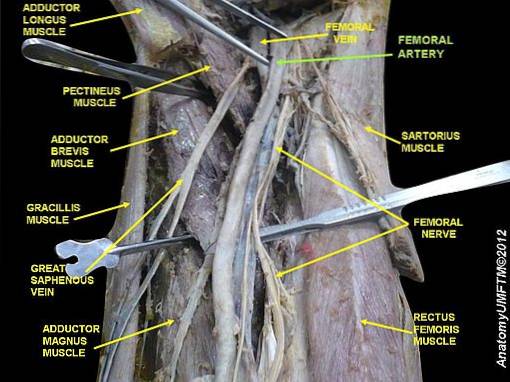
In its upper part it is located parallel to the femoral vein, but in an external position with respect to it. Descending distally, the femoral artery is anterior to the femoral vein. In its downward trajectory it is covered by the sartorius muscle.
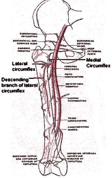
In addition to giving rise to the branches that supply the neighboring muscles and the skin, the femoral artery gives rise to 6 collateral branches which are:
1) Abdominal subcutaneous artery or epigastric artery superficialis.
2) Superficial circumflex iliac artery or circumflex artery ilium superficialis.
3) External pudendal arteries or external pudendal arteries.
4) Inguinal branches or groin rami.
5) Greater anastomotic artery or genus descendens artery.
6) Deep femoral artery or deep femoris artery.
In turn, the deep femoral artery gives rise to the internal circumflex artery with its two branches: the superficial and the deep, and the external circumflex artery with its ascending and descending branches. It also generates three perforating branches.
The femoral artery supplies the lower abdominal wall, the external genitalia and the lower limb, the upper thigh and, with its popliteal extension, supplies the knee, leg, and foot..
The arterial irrigation of the tissues brings nutrients and oxygen, which allows maintaining their metabolism and collecting, through the venous system, metabolic waste and CO2.
The branches of the femoral artery, as indicated above, are 6, then the route of each one and its irrigation areas will be defined.
It arises below the femoral arch, passes over the edge of the fascia lata and takes an upward path towards the umbilical region. It gives collateral branches that supply the skin and the greater oblique muscle of the abdomen..
It often presents as a branch of the abdominal subcutaneous artery, but in other cases it is a branch of the femoral artery. It is the smallest branch of the femoral artery.
It follows a superficial path over the fascia lata and goes towards the anterior superior iliac spine. Irrigate the skin, superficial fascia, and superficial groin nodes.
There are two or three arteries. Pass in front of or behind the femoral vein and irrigate the scrotum and penis in men, as well as the labia majora in women
They are branches that end in the lymph nodes and muscles of the triangle of Scarpa (anatomical space in the groin area).
It arises when the femoral artery passes through the canal of the adductors, perforates the canal in its anterior wall and descends, sliding through the sartorius muscle, passing behind the internal tuberosity of the femur..
It accompanies the saphenous nerve for a variable course. It supplies articular branches that help to form the joint network that irrigates the knee and muscular branches for the irrigation of the vastus medialis.
It is born two to six centimeters below the femoral arch and descends behind and outside the femoral artery that gave rise to it. It runs in front of the adductor medius, pectineus, and iliopsoas muscles. The median adductor covers it in its descent.
This artery has five main branches:
1) The internal circumflex artery
2) The external circumflex artery
3) Three perforating arteries
The first arises immediately below the origin of the internal femoral artery and passes behind the femoral arteries and vein on its descent. It gives rise to the superficial branch and the deep branch. They irrigate part of the hip joint, the skin and neighboring muscles such as the adductors or the pectineum, among others..
The second is born just in front of the anterior position and passes over the iliac psoas giving, in turn, two branches: the ascending one that supplies the tensor fascia lata and the gluteus, and the descending one that supplies the vastus external and femoral muscles. reaches the knee and irrigates the skin.
The perforating arteries supply the adductors as well as the skin and muscles of the posterior or dorsal thigh. The second perforator gives rise to the feeding artery of the femur.
Due to their superficial location within the femoral triangle in the groin, both the femoral artery and the femoral vein are vulnerable to lacerations, especially in anterior superior thigh injuries..
In these cases, as these vessels are quite thick and with a high flow rate, an injury that breaks these vessels can be fatal. This is because the blood loss is violent and very abundant, rapidly causing hypotension, loss of consciousness and death in a few minutes..
Atherosclerosis, which is a peripheral vascular disease in which atheromatous plaques accumulate on the internal surface of the arteries, can affect the femoral artery, generating, in some cases, occlusion of the arterial lumen.
Femoral occlusion is associated with severe non-irrigated or insufficiently irrigated limb pain, intermittent claudication, and cramps. Pain increases with exercise or movement and decreases with rest, but does not go away.


Yet No Comments