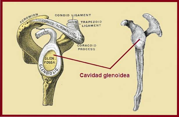
The glenoid cavity It is a concavity of the shoulder blade or scapula. The head of the humerus fits perfectly into it, forming the glenohumeral joint or also called the scapulohumeral joint. This joint is also known as the first shoulder joint..
This joint is very unstable and, therefore, the function of the deltoid muscle is to raise the humeral head towards the acromion, while the supraspinatus muscle does not allow the head of the humerus to leave the glenoid cavity.
In turn, in the middle arches of movement of the shoulder, the rotator cuff acts as a dynamic stabilizer, which is responsible for pushing the humeral head against the glenoid cavity.
Similarly, shoulder movements above 60 ° and in rotation, the joint is stabilized by the inferior glenohumeral complex. This complex is formed by the joint capsule in conjunction with the inferior glenohumeral ligament.
Among the pathologies that involve the glenoid cavity are: the instability of the shoulder whose cause is multifactorial, the osteoarthritis of the glenohumeral joint, fracture of the glenoid cavity and the Bankart lesion, among others..
Article index
The glenoid cavity is a shallow concavity, pear-shaped, being longer than it is wide, with a broader base.
According to Romero et al., The average measurement of the glenoid cavity in the cephalo-caudal area is 3.70 cm and the antero-posterior diameter is approximately 2.71 cm..
These data coincide with those obtained by Kose et al in 2018, who evaluated 100 patients, whose average cephalo-caudal area was 38.15 mm for the dominant side and 37.87 mm for the non-dominant side, while the anteroposterior diameter was 28, 60 mm for the dominant side and 28.00 mm for the non-dominant side.
This means that both glenoid cavities are not the same, and there are significant differences between them..
This information can be very useful in total shoulder prosthetic replacements, especially to correct current problems of loosening of the glenoid prosthetic device and the consequent glenohumeral instability..
On the other hand, the glenoid cavity has a ring of fibrocartilaginous tissue called the labrum or glenoid rim. The labrum, together with the joint capsule and the glenohumeral ligaments, is called the capsulolabral complex. Allows the concavity to be a little deeper, thus providing stability to the glenohumeral joint.
The shallow depth of the glenoid cavity gives it an advantage over the rest of the joints, since it allows the shoulder to have a fairly wide range of motion, being the joint that has the greatest capacity for movement. However, this same characteristic provides it with a disadvantage, since it makes it more vulnerable to suffering dislocations..
Its main function is to permanently receive and accommodate the head of the humerus, offering it the ability to move. Therefore, it is not a static relationship, but on the contrary, it is very dynamic.
It also serves as an insertion point for certain muscles, such as: the long head of the biceps is fixed on the upper edge of the glenoid cavity and the long head of the triceps that rests on the lower edge of the same socket.
Glenohumeral joint instability can be caused by: injury to the capsulolabral complex, excessive glean anteversion, or capsular hypermobility. On the other hand, there are studies that show that there are anatomical factors that can influence to increase the predisposition to have an unstable shoulder.
The anatomical parameters that are relevant in this regard are: the horizontal glenohumeral index, the glenoid inclination and the angle of anteversion of the scapula..
Glenohumeral joint instability can begin with a subluxation and end with a complete dislocation. This affectation is very frequent, it represents 95% of all dislocations, being more common in men than in women..
It should be noted that an unstable shoulder causes pain, limiting certain movements.
Treatment for glenoid cavity instability 100% surgical, as long as the number of dislocations is above three episodes.
The options are, the placement of special prostheses or the osteosynthesis or reconstruction of the fractures of the glenoid cavity.
To detect instability of the glenohumeral joint, several tests can be performed on the patient:
It is a rather uncomfortable test for the patient. An attempt is made to place the arm in abduction at 90 °, while inducing external rotation in retropulsion.
The patient's feeling under this action is that the shoulder will be dislocated, that is, he feels that the head of the humerus is going to come out of the glenoid cavity and of course he resists this movement.
With the patient lying on his back, leaving the shoulder to be examined off the table, the patient's arm is placed in external rotation and 90 ° abduction. From this position we place the hand behind the patient's shoulder and, as the rotation is increased, the shoulder is pushed forward.
As soon as the patient complains of pain, the opposite shoulder is pushed, that is, backwards. If this action causes the pain to be minimized or disappear, it is considered a positive test for glenohumeral instability..
This test assesses anteroposterior laxity. With the patient seated, the patient is asked to fully extend the arm to the side of the body, then the shoulder is stabilized and, with great care, an attempt is made to move the head of the humerus backwards and subsequently forwards..
The professional who performs the exam will be able to detect whether the movement of the shoulder is normal or abnormal..
Evaluates inferior instability of the glenohumeral joint. For this test the patient must be seated. You are asked to extend the arm to the side of the body and then to flex the elbow.
Starting from this position, a downward pull is performed. If it is possible to detect a depression below the acromion, it is a sign that there is a lesion of the rotator interval, and in this case the test is considered positive.
All imaging studies are valuable and each provides useful information, that is, they are complementary.
In this sense, radiology and Computerized Axial Tomography (CT) or arthro CT, offer precise information on bone lesions and guide towards the type of surgical treatment to be followed..
Whereas, magnetic resonance imaging is useful to study soft tissues, as for example in the case of a tear of the fibrocartilaginous tissue (labrum).
It is usually the result of a fracture. It begins with a non-surgical treatment and if it does not resolve, you should go to surgery. These pre-surgical options include arthrodesis or total or inverted prosthesis..
They are caused by trauma. This type of fracture requires surgical intervention, given its complexity. Idelberg classifies glenoid fractures into six categories according to the characteristics of the injury, such as extension of the fracture, structures involved or orientation of the fracture, among others..
The Bankart lesion is characterized by damage to the connective tissue that surrounds the glenoid cavity which, as we mentioned earlier, is called the labrum or glenoid rim.
It usually occurs after trauma, such as a shoulder dislocation. It is also possible that it will tear from repetitive movements during a sport. Glenoid labrum tear causes joint instability.
In this situation, the patient feels that the shoulder is going to slip out of place, in fact, it is possible. Also, the patient feels pain when moving the shoulder. In these cases, magnetic resonance imaging is ideal for making the diagnosis..
In mild injuries it is possible to treat with physiotherapy, but in more severe cases surgery is necessary.


Yet No Comments