The enterochromaffin cells, They are a type of intestinal endocrine and neuroendocrine cells. They are located next to the epithelium that lines the lumen of the digestive tract and affect a variety of physiological states.
Also known as ECL cells, they play a crucial role in gastrointestinal regulation, specifically in intestinal motility and secretion, in nausea and abdominal pain.
Most of the mechanical stimuli within the intestinal lumen do not interact directly with the afferent nerves, but instead activate specialized cells in the epithelium in a process of sensory transduction..
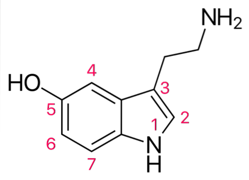
One of the first steps in this process is believed to be the release of the neurotransmitter biogenic serotonin amine (5-HT) by enterochromaffin cells.
Dietary nutrients and irritants, as well as gut-dwelling bacteria products and inflammatory agents, act on the intestinal epithelium to modulate signaling pathways that control digestion, immunity, metabolism, and pain.
Article index
Enterochromaffin cells comprise the major population of intestinal endocrine cells and play a critical role in various aspects of intestinal function including secretion, motility, and sensation..
They are responsible for the synthesis, storage and release of the largest store of 5-HT in the body. Produce more than 90% of the body's total serotonin, as well as a variety of peptides.
The synthesized serotonin is accumulated in secretory vesicles and uses a vesicular transporter called monoamine 1. In these secretory vesicles, serotonin is localized together with acidic proteins called chromogranins.
These vesicles fulfill various functions such as storage of proteins, amines and pro-hormones in cells..
The structure of most enterochromaffin cells is of the “open” type, that is, they present apical cytoplasmic extensions that project into the lumen of the gland with short microvilli, which favor the cellular response to physical or chemical variations..
It is believed that they also activate the mucosal processes of primary afferent neurons, through the release of serotonin from storage granules located at the base of cells..
Secreted serotonin can also influence neighboring cells (paracrine action). It also exerts a hormonal effect on distant cells through blood circulation.
Historically, various techniques have been used to visualize enterochromaffin cells..
In 1870, Heidenhain described these cells in the intestine and named them chromaffin cells, for their ability to stain brown when treated with chromic salts. Later, Kultschitzky described them as acidophilic basigranular cells..
Such cells can be identified by staining with chromium and silver salts and are therefore called enterochromaffin cells, which refers to their affinity for silver salts..
Today, more precise, reproducible and specific methods are used for the visualization and identification of enterochromaffin cells, such as staining techniques that use antibodies directed against serotonin..
In formalin-fixed intestinal mucosa tissues, it has been shown that enterochromaffin cells have very long and thin extensions that cross the connective tissue and neighboring glands..
They are small polygonal cells located in the crypts, between the intestinal villi. They present granules located in the basal region and containing serotonin and other peptides.
From a structural point of view, these granules have been reported to vary in size and shape..
The tissue beneath the enterochromaffin cells generally contains abundant fenestrated capillaries, lymphatic vessels, and small nerve fibers lacking myelin..
It is caused by the secretion of serotonin, dopamine, and catecholamines. Symptoms include diarrhea, abdominal cramps, flushing, sweating, and valvular heart disease..
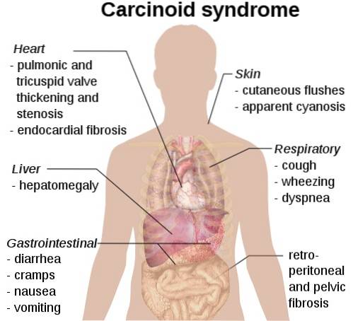
Excess circulating serotonin is generally produced by carcinoid tumors originating from enterochromaffin cells in the small intestine or appendix. They can also be present in other sites, particularly the lung and stomach.
This disease describes the cardiac and vascular changes associated with carcinoid syndrome. Fibrous plaques develop on the surface of the membrane that lines the inside of the heart's chambers (endocardium).
Plaques contain deposits of myofibroblasts, connective tissue cells, and smooth muscle cells.
The cause of carcinoid heart disease is not yet clear, however it has been suggested that serotonin is a possible agent involved in this pathogenesis..
This is a condition that involves chronic intestinal discomfort and abdominal pain. In this case, abnormal levels of serotonin have also been shown to be associated with this syndrome..
Irritable bowel syndrome can become severe and lead to chronic constipation or chronic diarrhea. Abnormal populations of enterochromaffin cells have been correlated with both conditions.


Yet No Comments